NeuroDex Contributes to Landmark Review on Tissue-Specific Extracellular Vesicles and the Future of Liquid Biopsy
February 2026 | NeuroDex, Inc.
NeuroDex is proud to announce the publication of a comprehensive peer-reviewed review article in the Journal of Extracellular Biology titled “Tissue-Specific Extracellular Vesicles Enriched From Circulation: Exploring the Liquid Biopsy Perspective.” This work reflects a major collaborative effort between academic and industry researchers and highlights NeuroDex’s continued commitment to advancing the science and clinical translation of extracellular vesicle (EV)-based biomarkers.
The publication explores one of the most critical challenges in modern diagnostics and precision medicine: how to reliably identify, enrich, validate, and apply tissue-specific extracellular vesicles (TS-EVs) as minimally invasive biomarkers for disease detection, progression monitoring, and therapeutic development. With contributors from leading institutions including Columbia University, Johns Hopkins University, and Harvard T.H. Chan School of Public Health, and with NeuroDex as an industry partner, the paper provides a roadmap for how EV science can move from exploratory research into standardized, reproducible, and clinically actionable liquid biopsy applications.
Understanding Extracellular Vesicles: Nature’s Biological Messengers
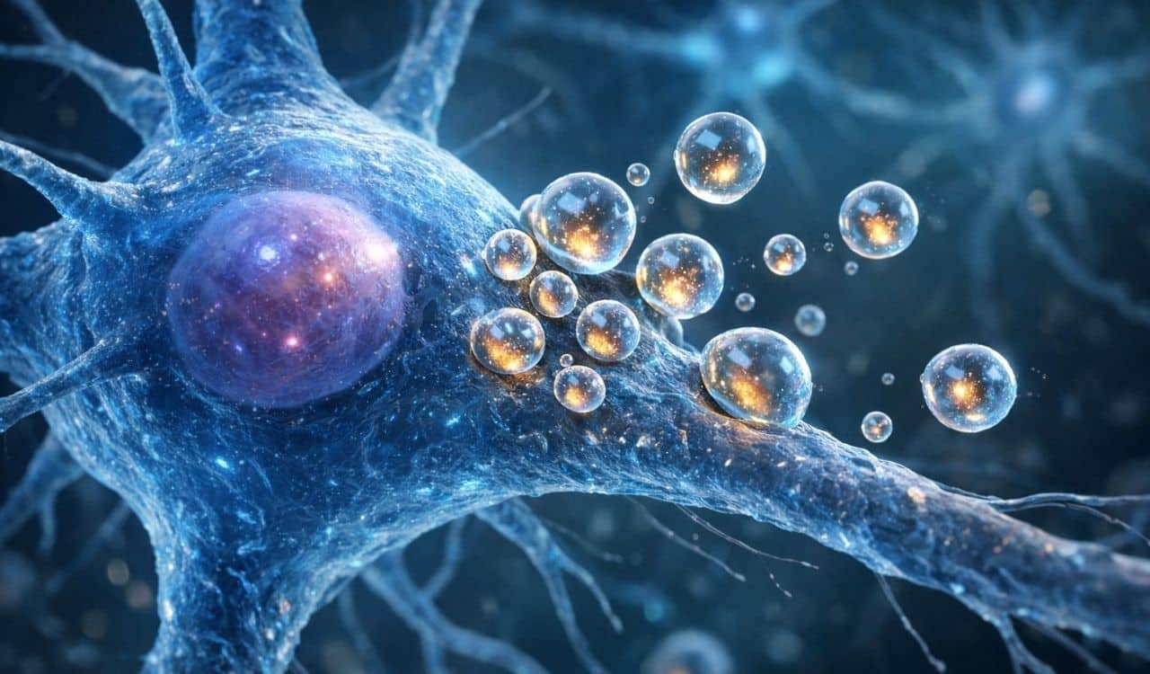
Extracellular vesicles are small, membrane-bound particles released by virtually all known cell types. Found in biofluids such as blood, cerebrospinal fluid, urine, saliva, and breast milk, EVs serve as critical mediators of intercellular communication. They carry a complex and highly informative molecular cargo that includes proteins, lipids, DNA fragments, messenger RNA, microRNAs, and other small molecules.
What makes EVs particularly powerful as biomarkers is their ability to reflect the phenotypic and functional state of their cells of origin. Under both healthy and pathological conditions, cells selectively package molecular signals into EVs, which are then released into local tissue environments or into systemic circulation. These vesicles can cross biological barriers, including the blood–brain barrier, allowing tissues that are otherwise difficult or risky to biopsy to be studied through a simple blood draw.
In recent years, EVs have emerged as a promising foundation for liquid biopsy, a minimally invasive diagnostic approach that enables clinicians and researchers to detect molecular signatures of disease without the need for surgical tissue sampling. While liquid biopsy has traditionally relied on circulating tumor DNA and circulating tumor cells, EVs offer a complementary and in many cases more tissue-specific window into disease biology.
The Challenge: Complexity and Heterogeneity in Circulating EVs
Despite their promise, EVs present significant scientific and technical challenges. Blood and other biofluids contain billions of vesicles per milliliter, originating from a wide range of tissues and cell types. The majority of circulating EVs are derived from blood cells, such as platelets and other hematopoietic sources. In contrast, EVs originating from organs like the brain, liver, heart, lungs, or placenta typically represent only a small fraction of the total circulating population.
This creates a fundamental problem for biomarker discovery: the signals of interest can be overwhelmed by background noise. A disease-related molecular marker carried by EVs from a specific tissue may be diluted or obscured when analyzing total EV populations. As a result, the field has increasingly focused on methods to selectively enrich and analyze tissue-specific EV subpopulations.
The review published by NeuroDex and its collaborators systematically examines the biological basis of EV heterogeneity and the technical strategies currently used to address it. The authors highlight that EV diversity arises from multiple dimensions, including:
Differences in cellular origin
Variations in biogenesis pathways
Diversity in surface protein composition
Differences in size, density, and cargo content
This heterogeneity, while biologically meaningful, complicates efforts to develop standardized and reproducible diagnostic workflows.
Tissue-Specific EVs: A New Frontier in Liquid Biopsy
Tissue-specific EVs represent a subset of circulating vesicles that carry surface markers and molecular cargo uniquely associated with a particular tissue or cell type. By targeting these features, researchers can isolate EVs originating from organs such as the brain, liver, heart, adipose tissue, placenta, or lungs.
The review outlines how TS-EVs can serve as minimally invasive surrogates for tissue health, enabling:
Early disease detection
Monitoring of disease progression
Assessment of therapeutic response
Discovery of new molecular targets
This approach is especially valuable for tissues that are difficult to access through conventional biopsy methods. For example, obtaining brain tissue from living patients is rarely feasible, yet neuronal EVs circulating in blood can carry proteins and nucleic acids reflective of neurodegenerative processes.
NeuroDex’s Focus: EVs in Neurodegenerative Disease
At NeuroDex, EV-based liquid biopsy is central to the company’s mission of transforming the diagnosis and development of treatments for neurodegenerative diseases. Conditions such as Alzheimer’s disease, Parkinson’s disease, amyotrophic lateral sclerosis (ALS), and frontotemporal dementia are characterized by complex and often silent pathological processes that begin years before clinical symptoms appear.
The review highlights extensive research into neuron-derived extracellular vesicles, which have been shown to carry hallmark proteins associated with neurodegeneration, including misfolded or pathological forms of tau, alpha-synuclein, amyloid-beta, and TDP-43. These molecules are central to disease mechanisms and are actively being investigated as diagnostic and prognostic biomarkers.
NeuroDex’s involvement in this publication reflects its commitment to advancing:
Robust methods for neuronal EV enrichment
Multi-marker validation strategies
Integration of proteomic and transcriptomic analysis
Translational workflows aligned with clinical and regulatory requirements
By contributing to the broader scientific dialogue on validation and standardization, NeuroDex aims to help establish EV-based diagnostics as reliable tools for clinical decision-making.
Methods for Enriching Tissue-Specific EVs
The review provides a detailed overview of current strategies used to isolate and characterize TS-EVs from complex biological fluids. These methods fall into two broad categories: physical property-based separation and immunoaffinity-based capture.
Physical Separation Techniques
These approaches rely on differences in EV size, density, or charge. Common techniques include:
Ultracentrifugation
Density gradient centrifugation
Size exclusion chromatography
Ultrafiltration
Ion exchange chromatography
While these methods are effective for enriching total EV populations, they generally lack the specificity needed to isolate vesicles from a particular tissue. They may also co-isolate other extracellular particles, such as lipoproteins and protein aggregates, which can interfere with downstream analyses.
Immunoaffinity-Based Capture
Immunoaffinity methods target surface proteins expressed on EV membranes that reflect the identity of their parental cells. By using antibodies or other binding molecules attached to magnetic beads, microfluidic devices, or solid supports, researchers can selectively isolate EV subpopulations of interest.
The review highlights this approach as one of the most promising pathways toward true tissue-specific liquid biopsy. However, it also underscores the need for careful validation, as surface markers may not be exclusively expressed by a single tissue, and soluble forms of some proteins can lead to non-specific binding.
The Importance of Validation and Reproducibility
One of the central themes of the publication is the urgent need for methodological rigor. Across the field, studies often use different protocols, markers, and analytical workflows, making it difficult to compare results or reproduce findings.
The authors identify several key gaps that must be addressed for EV-based diagnostics to reach clinical maturity:
Limited use of isotype controls to assess non-specific binding
Incomplete validation of tissue specificity
Lack of standardized efficiency and recovery metrics
Insufficient reporting of protocol details
Without these elements, even promising biomarker discoveries may fail to translate into reliable clinical tests. The review calls for the adoption of multi-marker validation frameworks, where both EV surface markers and internal cargo are analyzed to confirm tissue origin.
Applications Across Organ Systems
The publication explores the role of TS-EVs across a wide range of tissues and disease contexts, demonstrating the broad relevance of this technology.
Nervous System
Neuronal, astrocytic, microglial, and oligodendrocyte-derived EVs have been investigated as biomarkers for neurodegenerative and neuroinflammatory disorders. These vesicles can cross the blood–brain barrier, making them uniquely valuable for studying central nervous system pathology through peripheral blood samples.
Liver
Hepatocyte-derived EVs have been linked to metabolic disorders, fibrosis, and liver cancer. Specific surface markers and cargo molecules have shown promise in distinguishing between different stages of liver disease.
Heart
Cardiomyocyte-derived EVs are being explored as indicators of myocardial stress, injury, and remodeling. Emerging studies suggest these vesicles may provide insight into pathways involved in heart failure and ischemic injury.
Adipose Tissue
Adipocyte-derived EVs play a role in systemic metabolic regulation and inflammation. They have been associated with obesity, insulin resistance, and cardiovascular risk.
Placenta
Placenta-derived EVs contribute to maternal–fetal communication and have potential applications in monitoring pregnancy-related conditions such as pre-eclampsia and gestational diabetes.
Microbial EVs
The review also discusses bacterial-derived vesicles, which influence host immune responses and may serve as biomarkers of microbiome-related disease processes.
Implications for Precision Medicine
The ability to non-invasively access tissue-specific molecular information has profound implications for precision medicine. EV-based liquid biopsy has the potential to:
Enable earlier diagnosis of disease
Improve patient stratification for clinical trials
Monitor therapeutic response in real time
Reduce reliance on invasive procedures
By capturing dynamic molecular signals, TS-EVs offer a longitudinal view of disease biology that is difficult to achieve with traditional tissue biopsies.
For pharmaceutical and biotechnology partners, this approach can enhance drug development by providing biomarkers that reflect target engagement, disease modulation, and safety profiles.
NeuroDex’s Translational Vision
NeuroDex is dedicated to bridging the gap between cutting-edge EV research and real-world clinical application. The company’s platform integrates:
Standardized EV enrichment workflows
Multi-omic analysis pipelines
Clinical laboratory quality systems
Regulatory-aligned validation frameworks
Through partnerships with academic institutions, clinical centers, and industry collaborators, NeuroDex aims to accelerate the development of EV-based diagnostics and biomarker services that can support both clinical care and therapeutic innovation.
This publication represents an important step in that journey, reinforcing NeuroDex’s role in shaping the scientific and methodological foundations of EV-based liquid biopsy.
Looking Ahead: Building the Future of EV Diagnostics
The review concludes with a forward-looking perspective on the evolution of tissue-specific EV research. While significant progress has been made, the field is still in an early phase of development. Achieving widespread clinical adoption will require:
Community-driven standardization efforts
Cross-laboratory validation studies
Regulatory engagement
Integration with clinical workflows
NeuroDex is committed to contributing to these efforts by supporting open scientific dialogue, collaborative research, and the development of robust, scalable diagnostic platforms.
Read the Full Publication
The full open-access review article is available at:
https://doi.org/10.1002/jex2.70106
About NeuroDex
NeuroDex is a biotechnology company focused on advancing liquid biopsy diagnostics and biomarker discovery through tissue-specific and neuron-derived extracellular vesicle technologies. The company partners with academic, clinical, and industry leaders to enable precision medicine approaches in neurodegenerative and complex diseases.
For more information, visit:
This article is intended for informational purposes only and does not constitute medical or regulatory advice.


Leave a Reply