Extracellular Vesicles in Neuroscience: A Definitive Guide for Researchers
Introduction: The Emerging Role of Extracellular Vesicles in Brain Communication
The intricate landscape of the brain, long understood through the lens of neuronal electrical signaling and synaptic transmission, is revealing a far more complex communication network. At the forefront of this revelation are extracellular vesicles (EVs), tiny membrane-bound sacs released by virtually all cells, including those in the central nervous system (CNS). Once dismissed as cellular debris, EVs are now recognized as sophisticated messengers, carrying a diverse cargo of biomolecules that profoundly influence cellular function. For researchers in neuroscience, understanding EVs is no longer an option but a necessity, offering unprecedented insights into healthy brain function, the pathogenesis of devastating neurological disorders, and novel therapeutic avenues. This guide aims to provide a definitive overview of EVs, their roles in neuroscience, and the cutting-edge methodologies used to study them.
1 What are Extracellular Vesicles (EVs)?
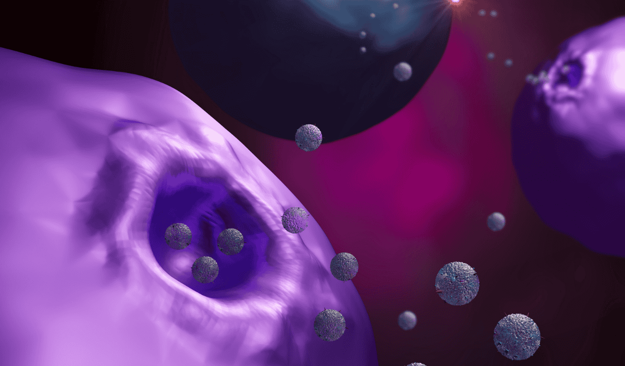
![]() The two primary pathways of EV biogenesis: exosomes are formed within the endosomal system and released upon fusion with the cell membrane, while microvesicles bud directly from the cell surface.
The two primary pathways of EV biogenesis: exosomes are formed within the endosomal system and released upon fusion with the cell membrane, while microvesicles bud directly from the cell surface.
Extracellular vesicles (EVs) are nano- to micro-sized particles secreted by cells into the extracellular environment. They are distinct from apoptotic bodies and are broadly classified into two main categories based on their biogenesis: exosomes and microvesicles. Exosomes are typically smaller (30-150 nm) and originate from the endosomal pathway, forming within multivesicular bodies (MVBs) before fusing with the plasma membrane to be released. Microvesicles, also known as ectosomes, are generally larger (50-1000 nm) and are formed by direct outward budding of the plasma membrane. Both types of vesicles are enclosed by a lipid bilayer and serve as crucial vehicles for intercellular communication, transporting a rich content of proteins, lipids, and nucleic acids between cells. The sheer number of these vesicles in biological fluids like cerebrospinal fluid (CSF) and blood underscores their widespread involvement in physiological and pathological processes within the neuroscience domain.
2 Why EVs Matter in Neuroscience
The brain is an exceptionally complex organ with a high metabolic rate and a critical need for precise intercellular communication. EVs play a pivotal role in this intricate network. They are released by neurons, glial cells (astrocytes, microglia, oligodendrocytes), and endothelial cells of the blood-brain barrier, acting as messengers that can influence nearly every aspect of brain function. This includes modulating synaptic plasticity, promoting neuronal survival, facilitating myelination, and orchestrating immune responses within the CNS. Moreover, EVs can traverse biological barriers, including the formidable blood-brain barrier (BBB), making them potent mediators of communication between the brain and the periphery. Understanding their roles is thus fundamental to deciphering normal brain physiology and uncovering the mechanisms underlying neurological diseases. The ability of EVs to carry specific information and support cellular processes makes them central players in maintaining brain health.
EV Biogenesis, Cargo, and Release: Understanding the Molecular Machinery
The functional diversity of extracellular vesicles stems directly from the complex processes governing their formation, the composition of their internal cargo, and the mechanisms by which they are released from parent cells. A thorough understanding of these cellular events is critical for appreciating how EVs exert their influence in neuroscience.
1 Diverse Origins and Biogenesis Pathways
The distinct biogenesis pathways of exosomes and microvesicles dictate their physical properties and, to some extent, their cargo and function. Exosome formation is intricately linked to the endosomal sorting complexes required for transport (ESCRT) machinery, although ESCRT-independent pathways also exist. Intraluminal vesicles (ILVs) form within late endosomes, creating multivesicular bodies (MVBs). These MVBs can then follow one of two fates: fusion with lysosomes for degradation or fusion with the plasma membrane, releasing the enclosed ILVs as exosomes into the extracellular space. Microvesicles, conversely, are generated by direct fission from the plasma membrane. This process involves the outward budding of the cell membrane, driven by cytoskeletal rearrangements and lipid domain organization. The specific cellular context and the signaling cascades active during vesicle formation heavily influence the composition of the resulting vesicles.
2 The Information Payload: A Rich Cargo of Biomolecules
The true power of extracellular vesicles lies in their content, which is a carefully selected and packaged repertoire of biomolecules, acting as a “message in a bottle” to recipient cells. This cargo includes a wide array of proteins, such as enzymes, receptors, transcription factors, and structural proteins. The lipids that form the EV membrane also play crucial roles, influencing membrane fluidity and facilitating interactions with target cells. Crucially, EVs encapsulate nucleic acids, including messenger RNAs (mRNAs), microRNAs (miRNAs), and even DNA fragments. These nucleic acids can be translated into proteins or regulate gene expression in recipient cells, profoundly impacting their function, differentiation, and survival. This diverse molecular payload allows EVs to mediate complex cellular dialogues, transferring critical information that can modify the behavior of neighboring or distant cells. The number of different molecules carried by a single EV can range from hundreds to thousands, highlighting their capacity as sophisticated carriers of cellular content.
Extracellular Vesicles in Healthy Brain Function: Orchestrating Neural Communication
In a healthy brain, extracellular vesicles are not passive bystanders but active participants in the dynamic interplay between neural and glial cells. They contribute to the maintenance of homeostasis, synaptic plasticity, and overall neuronal health, acting as crucial conduits for intercellular information exchange.
1 EVs as Mediators of Cell-to-Cell Communication in the CNS
Within the central nervous system, EVs facilitate communication between neurons, astrocytes, microglia, and oligodendrocytes. Neuronal EVs can influence synaptic transmission and plasticity, potentially delivering factors that enhance or suppress neurotransmitter release, or modulating the expression of synaptic proteins in recipient neurons. Astrocytes release EVs that can affect neuronal excitability and glial cell behavior, playing roles in neuroinflammation and repair. Microglia, the resident immune cells of the brain, also release EVs that can modulate neuronal function and communicate with other glial cells. Oligodendrocytes utilize EVs for myelin maintenance and repair. These interactions highlight how EVs support the complex communication networks essential for cognitive function, learning, and memory. The diverse content of these EVs, tailored by their cell of origin, ensures specific and targeted signaling.
2 Navigating Biological Barriers: The Blood-Brain Barrier
The blood-brain barrier (BBB) is a highly selective semipermeable border that protects the brain from circulating pathogens and toxins, but it also poses a significant challenge for therapeutic interventions. Extracellular vesicles, however, possess unique properties that allow them to navigate this barrier. EVs can be released from endothelial cells of the BBB, influencing its integrity and function. Conversely, EVs circulating in the blood can cross the BBB, and EVs released by brain cells can enter the circulation. This bidirectional trafficking of EVs allows for a continuous exchange of molecular information between the CNS and the periphery. This capacity is particularly relevant for understanding systemic influences on brain health and disease, as well as for developing diagnostic tools and therapies that can reach the brain. This ability to overcome such a formidable barrier is a key reason why EVs are of such immense interest in neuroscience.
EVs in Neurodegenerative Diseases: Insights into Pathogenesis and Progression
The aberrant behavior and dysregulation of extracellular vesicles are increasingly implicated in the pathogenesis and progression of a wide spectrum of neurodegenerative diseases. Their capacity to propagate misfolded proteins and inflammatory signals makes them central to understanding how these debilitating conditions develop and spread throughout the brain.
1 Alzheimer’s Disease (AD): EV-Mediated Spread of Pathology
In Alzheimer’s disease (AD), EVs play a critical role in the propagation of key pathological hallmarks: amyloid-beta (Aβ) plaques and hyperphosphorylated tau tangles. Neurons and other brain cells release EVs containing Aβ peptides and tau proteins. These EVs can then deliver these toxic species to neighboring cells, seeding the aggregation and spread of pathology throughout the brain. For instance, EVs released by AD-affected neurons have been shown to deliver tau seeds to healthy neurons, initiating tau pathology in those cells. The content of these EVs, including specific nucleic acids and modified proteins, can prime recipient cells for further aggregation and neurotoxicity. Understanding this EV-mediated spread is crucial for developing strategies to halt or slow the progression of AD.
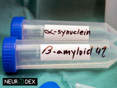
2 Parkinson Disease (PD): Alpha-Synuclein Propagation
Similar to AD, Parkinson disease (PD) is characterized by the accumulation of misfolded proteins, most notably alpha-synuclein, forming Lewy bodies. Extracellular vesicles are recognized as key players in the cell-to-cell transmission of alpha-synuclein pathology in PD. Neurons affected by PD release EVs containing alpha-synuclein monomers and oligomers. These EVs can then be taken up by healthy neurons, triggering the misfolding and aggregation of alpha-synuclein within the new host cell, thereby propagating the disease process. The presence of specific proteins and nucleic acids within these EVs can prime recipient cells for aggregation, contributing to the progressive loss of dopaminergic neurons that defines PD.
3 Other Neurodegenerative Disorders
The role of EVs extends beyond AD and PD to encompass other neurodegenerative diseases, including Huntington’s disease (HD) and Amyotrophic Lateral Sclerosis (ALS). In HD, EVs can carry mutant huntingtin protein fragments, contributing to its spread and neurotoxicity. In ALS, EVs have been implicated in the spread of misfolded TDP-43 protein and in modulating motor neuron survival. Across these diverse conditions, EVs often serve as conduits for the transfer of misfolded proteins, toxic RNA species, and inflammatory mediators, contributing to a common theme of intercellular pathology spread. The specific content of these EVs varies depending on the disease, offering potential targets for diagnosis and therapy.
4 The Role of Aging in EV Function and Disease Susceptibility
Aging is the most significant risk factor for most neurodegenerative diseases. As we age, cellular processes, including EV biogenesis and function, undergo significant changes. Older individuals tend to release EVs with altered content, potentially containing more pro-inflammatory molecules or misfolded proteins. Furthermore, the cellular machinery responsible for clearing EVs and their pathological cargo may become less efficient with age. This age-related shift in EV dynamics can increase susceptibility to the development and progression of neurodegenerative conditions. The number and composition of EVs can change, altering their capacity to support or disrupt cellular homeostasis, thereby contributing to the increased incidence of brain pathology in older populations.
EVs in Other Neurological Conditions and Brain Injury
The influence of extracellular vesicles is not confined to neurodegenerative disorders. They are also implicated in a range of other neurological conditions, including psychiatric and neurodevelopmental disorders, and play crucial roles in the aftermath of brain injury.
1 Psychiatric Disorders: Schizophrenia and Bipolar Disorder
Emerging research suggests that EVs may contribute to the pathophysiology of psychiatric disorders like schizophrenia and bipolar disorder. Alterations in the content and function of EVs in the brains of individuals with these conditions have been observed, potentially affecting neurotransmitter signaling, synaptic plasticity, and neuroinflammation. For example, changes in the miRNA content of EVs might influence gene expression in neurons and glial cells, impacting mood regulation and cognitive function. The specific proteins carried by EVs could also mediate inflammatory pathways implicated in these disorders.
2 Neurodevelopmental Disorders: Autism Spectrum Disorder
In autism spectrum disorder (ASD), EVs are being investigated for their potential role in altered neural connectivity and synapse development. EVs released by neurons and glial cells could influence the formation and maturation of synaptic connections during critical developmental periods. Dysregulation in EV trafficking or content might contribute to the atypical sensory processing and social interaction patterns observed in ASD. Research is exploring whether specific EV biomarkers could aid in early diagnosis or understanding of ASD subtypes.
3 Brain Injury and Peripheral Nerve Regeneration
Following acute brain injury, such as traumatic brain injury (TBI) or stroke, extracellular vesicles play a dual role. Initially, they can contribute to secondary injury through the release of inflammatory factors. However, in the later stages, EVs, particularly those released by mesenchymal stem cells (MSCs), can promote repair and regeneration. They can deliver neurotrophic factors, anti-inflammatory molecules, and genetic material that support neuronal survival and axon growth. Muscle-derived EVs have also been shown to influence peripheral nerve regeneration, demonstrating their broad impact on neural repair mechanisms across different contexts.
Methodologies for EV Isolation, Characterization, and Cargo Analysis
The burgeoning field of EV research relies heavily on robust methodologies for isolating, characterizing, and analyzing the content of these tiny messengers. Advances in these techniques are crucial for unlocking their full potential in neuroscience.
1 EV Isolation Techniques: Achieving Purity and Specificity
Isolating pure and functional extracellular vesicles from complex biological matrices like brain tissue or biofluids is a significant challenge. Common methods include differential ultracentrifugation, size exclusion chromatography (SEC), precipitation kits, and immunoaffinity capture. Each technique has its advantages and limitations, and the choice often depends on the source material and the downstream application. For example, ultracentrifugation is a widely used method but can lead to contamination with protein aggregates or other cellular debris. SEC offers better separation based on size and is less denaturing. Immunoaffinity capture uses antibodies against EV surface markers to achieve high specificity but can be costly. The global exosome isolation service market, valued at approximately USD 112 million in 2023 and projected to reach USD 540 million by 2032, growing at a CAGR of 19.2%, reflects the significant and growing demand for efficient isolation techniques. This growth signifies the critical need for reproducible and scalable methods to obtain pure EV samples for research and clinical applications.
2 EV Characterization and Quantification: A Multifaceted Approach
Once isolated, EVs must be rigorously characterized to confirm their identity and quantify their number and properties. A multifaceted approach is essential, integrating various techniques. Electron microscopy (EM), including transmission electron microscopy (TEM) and scanning electron microscopy (SEM), is vital for visualizing the morphology and size of vesicles, confirming their cup-like or spherical shape. Flow cytometry offers a powerful tool for high-throughput phenotyping and quantification of EV populations, especially when combined with technologies like PICO (particle imaging flow cytometry) capable of detecting single EVs. Nanoparticle tracking analysis (NTA) provides real-time measurements of particle size distribution and concentration. Western blotting is commonly used to detect specific EV marker proteins (e.g., CD9, CD63, TSG101, Alix), which help confirm their origin and purity.
3 Cargo Analysis: Unveiling the Molecular Content
Analyzing the molecular content of EVs is key to understanding their functional role. Proteomics approaches can identify thousands of proteins within EV populations, revealing their origin and potential signaling capabilities. Lipidomic analyses provide insights into the lipid composition of the EV membrane, which influences their stability and interactions. Transcriptomic analysis, particularly of miRNAs and mRNAs, allows researchers to understand the genetic information being transferred. Techniques like mass spectrometry for proteomics and RNA sequencing for transcriptomics are indispensable for comprehensively characterizing the cargo of extracellular vesicles.
4 Ensuring Reproducibility and Standardization in EV Research
A significant challenge in EV research is achieving reproducibility and standardization across different laboratories and studies. The International Society for Extracellular Vesicles (ISEV) has published Minimum Information for Studies of Extracellular Vesicles (MISEV) guidelines to promote best practices for EV isolation, characterization, and data reporting. Adherence to these guidelines is crucial for comparing results and building a reliable body of knowledge. Ensuring the purity of isolated EVs, accurately quantifying their number, and consistently reporting their molecular content are paramount for advancing the field and translating EV research into clinical applications.
Extracellular Vesicles as Biomarkers: The Promise of Liquid Biopsy Diagnostics
The unique characteristics of extracellular vesicles position them as highly promising biomarkers for early diagnosis, prognosis, and monitoring of neurological diseases, offering the prospect of revolutionary liquid biopsy diagnostics.
1 Advantages of EVs in Biomarker Discovery
EVs offer several distinct advantages as biomarkers. They are present in virtually all bodily fluids, including blood and CSF, allowing for minimally invasive sample collection. The lipid bilayer of EVs protects their enclosed cargo from degradation, rendering them more stable in circulation than free biomolecules. Furthermore, EVs carry a diverse cargo reflective of their cell of origin, meaning that EV-derived biomarkers can potentially offer a more specific and sensitive window into pathological processes occurring within the brain. The content of EVs—specific proteins, nucleic acids, and lipids—can serve as highly informative disease signatures.
2 Biomarker Candidates in Cerebrospinal Fluid (CSF) and Blood
Numerous studies have identified EV-derived biomarkers for various neurological conditions. In Alzheimer’s disease, EVs isolated from CSF or blood have been shown to contain specific forms of tau proteins, amyloid-beta peptides, and dysregulated miRNAs that correlate with disease severity and progression. For Parkinson disease, EV-associated alpha-synuclein species and specific miRNAs are being investigated. The number and specific molecular signature of EVs can also change in response to aging and various neurological insults. Researchers are actively seeking robust EV biomarkers in both CSF and blood that can reliably distinguish between healthy individuals and those with neurological disorders.
3 Liquid Biopsy for Early Diagnosis and Monitoring
The development of liquid biopsy diagnostics based on extracellular vesicles holds immense potential for transforming patient care in neuroscience. The ability to detect subtle changes in EV cargo and number in blood or CSF at the earliest stages of disease could enable interventions before irreversible damage occurs. This approach is particularly valuable for conditions like AD and PD, where diagnosis is often delayed. EV-based liquid biopsies could also be used to monitor disease progression, therapeutic response, and disease recurrence, offering a more personalized and dynamic approach to managing neurological conditions. The North America exosomes market, accounting for 56.55% of revenue share in 2024, demonstrates significant investment in this area, driven by the promise of such diagnostic advancements.
Therapeutic Applications of Extracellular Vesicles
Beyond their diagnostic potential, extracellular vesicles are emerging as powerful therapeutic agents and delivery vehicles, offering novel strategies for treating neurological disorders and promoting tissue repair.
1 EVs as Natural Drug Delivery Vehicles
The inherent properties of EVs make them ideal natural drug delivery vehicles. Their biocompatibility and low immunogenicity reduce the risk of adverse reactions. Crucially, EVs can naturally cross biological barriers, including the blood-brain barrier, which remains a major hurdle for conventional therapeutics; only an estimated 2% of new CNS therapeutic compounds can cross the BBB and reach their therapeutic targets. Researchers are engineering EVs to encapsulate therapeutic payloads, such as small molecules, peptides, or nucleic acids (like siRNAs or miRNAs), to deliver them directly to target cells within the brain. The content of these engineered EVs can be tailored to achieve specific therapeutic effects, such as reducing inflammation, promoting neurogenesis, or delivering gene-editing machinery. The global extracellular vesicle therapy market size, valued at USD 750 million in 2023 and projected to grow to USD 1566 million by 2030, with a CAGR of 11.3%, underscores the significant investment and anticipation surrounding EV-based therapeutics.
Conclusion
The journey of extracellular vesicles from cellular obscurity to the vanguard of neuroscience research is a testament to their profound biological significance. These tiny vesicles are far more than mere waste products; they are sophisticated communication agents, carrying vital information in the form of proteins, lipids, and nucleic acids that orchestrate complex cellular interactions within the brain. From maintaining healthy neural function to driving the pathology of neurodegenerative diseases like Alzheimer’s disease, their influence is pervasive. The advancements in isolation and characterization techniques, including flow cytometry and electron microscopy, are continuously refining our ability to study them. Their potential as non-invasive biomarkers for liquid biopsy diagnostics promises to revolutionize early detection and monitoring, while their capacity as natural drug delivery vehicles opens new frontiers in therapeutic intervention. As research progresses, a deeper understanding of EV biogenesis, cargo selection, and release mechanisms, alongside concerted efforts towards standardization, will be crucial. The increasing number of studies and the robust growth in the EV market, with projections indicating significant expansion, underscore the transformative potential of EVs. By harnessing the power of EVs, researchers are poised to unlock unprecedented insights into brain function and develop innovative strategies to combat neurological disorders, offering renewed hope for millions affected by these conditions.


Leave a Reply