
LuminEV Assay Manual
This package insert must be read in its entirety before using this product.
FOR RESEARCH USE ONLY. NOT FOR USE IN DIAGNOSTIC PROCEDURES.
Catalog numbers
• LX-tspn-01: 1-plate pack
• LX-tspn-05: 5-plate pack
• LX-tspn-20: 20-plate pack
Table of contents
Introduction………………………………………………………………………………………………………………………….. 2
Reagents supplied…………………………………………………………………………………………………………………. 2
Reagents not supplied…………………………………………………………………………………………………………… 3
Assay principle………………………………………………………………………………………………………………………. 3
Safety precautions………………………………………………………………………………………………………………… 3
Materials required………………………………………………………………………………………………………………… 3
Equipment required………………………………………………………………………………………………………………. 4
Technical guidelines………………………………………………………………………………………………………………. 4
Reagent preparation……………………………………………………………………………………………………………… 5
Plate washing………………………………………………………………………………………………………………………… 6
Equipment settings……………………………………………………………………………………………………………….. 7
Assay protocol………………………………………………………………………………………………………………………. 8
Validation……………………………………………………………………………………………………………………………… 9
Analysis of results…………………………………………………………………………………………………………………. 9
Contact information
Customer service:
Email: customerservice@neurodex.co
Scientific support:
Email: scientificsupport@neurodex.co
Introduction
LuminEV enables the measurement of surface tetraspanins CD9, CD63, and CD81 on intact extracellular vesicles in unprocessed plasma, cell culture media, and other biological fluids.
Reagents supplied
| Reagent | Storage | Catalog No. | Quantity Supplied (Plates) | Description | ||
| 1 ea | 5 ea | 20 ea | ||||
| CD9 capture (40x) | 4C | C-01-tspn | 150 ul | 750 ul | 3000ul | Microsphere-conjugated capture antibody |
| Capture CD63 (40x) | 4C | C-02-tspn | 150 ul | 750 ul | 3000ul | Microsphere-conjugated capture antibody |
| Capture CD81 (40x) | 4C | C-03-tspn | 150 ul | 750 ul | 3000ul | Microsphere-conjugated capture antibody |
| Capture control (40x) | 4C | C-00-tspn | 150 ul | 750 ul | 3000ul | Microsphere-conjugated control antibody |
| Detection cocktail (200x) | 4C | D-01-tspn | 60 ul | 300 ul | 1200 ul | Biotinylated detection antibody cocktail |
| Standard (low TSPN content) | -80C | S-01-tspn | 0.25 mg | 1.25 mg | 5 mg | Purified EV from HEK293 culture media |
| Standard (high TSPN content) | -80C | S-02-tspn | 0.25 mg | 1.25 mg | 5 mg | Purified EV from U87mg culture media |
| Streptavidin-phycoerythrin (5x) | 4C | SAPE-01 | 1 ml | 5 ml | 20 ml | Reporter reagent for visualization of detection |
| Assay diluent (1x) | 4C | B-01-tspn | 20 ml | 100 ml | 500 ml | Diluent for capture, samples, standards, and detection antibodies |
Reagents not supplied
- Sheath fluid concentrate (Luminex, cat. 139005 or equivalent)
- PBS-T wash buffer powder (VWR, cat. 79371-736 or equivalent)
- DPBS (Gibco, cat. 10010-031 or equivalent)
- WFI (Gibco, cat. A12873-02 or equivalent)
Assay principle
This immunoassay assay employs the sandwich principle offers the capacity for multiplexing based on Luminex® technology. Capture antibodies are covalently linked to the color-coded magnetic microspheres with emission in distinct spectrum regions (capture beads). A mix of capture beads targeting distinct antigens are incubated with the sample, unbound material is washed away, and a bound material is detected with the biotinylated detection antibody of choice. The binding of the detection antibody is visualized with a streptavidin-phycoerythrin (SAPE) reagent. The microspheres are washed and re-suspended in sheath fluid. The assay is than read using the Luminex® instrument that detects capture beads with expected emission spectra and positive for PE, which is indicative for the presence of the analyte of interest.
Safety precautions
Use laboratory safety practices when handling assay components. Handle and dispose of biological materials in accordance with international, federal, state, and local regulations. Please note that sodium azide has been added to some reagents as a preservative, and thus, unused kit contents should be handled and disposed of properly.
Materials required
- F-bottom black plates (Brand Scientific, cat. 781608 or equivalent)
- 0.22um filter plate (Millipore Sigma, cat. MSGVS2210)
* Only required when using cell culture media.
- U-bottom dilution plate (Ratio Labs, cat. 6018113 or equivalent)
- Black vinyl plate sealers (Thermo Fisher, cat. 757-11047-CS or equivalent)
* Transparent plate sealers may be used if protected from light.
- 15ml black tubes (Celltreat, cat. 229431 or equivalent)
* Transparent tubes may be used if protected from light.
- Single Pipettes, suitable for dispensing 25-500ul
- Multichannel pipettes, suitable for dispensing 50-200ul
- Polypropylene microcentrifuge tubes
Equipment required
- Microplate spinner (Millipore Sigma, cat. Z742568 or equivalent)
- Ultrasonic water bath (Branson Sonifier, cat. CPX-952-116R or equivalent)
- Automatic plate washer for magnetic beads (BioRad cat. 30034376 or equivalent) or hand-held magnetic separator plate (Millipore Sigma, cat. Z740159 or equivalent)
- Mini tube rotator (Boekel Scientific, cat. 260750 or equivalent)
- Vortex mixer (Benchmark, cat. BV101 or equivalent)
- Plate shaker (Thermo Fisher cat. 88882005 or equivalent)
- Tube rockers (Thermo Fisher cat. 88882017 or equivalent)
- Luminex 200, HTS, Flexmap 3D, or MagPix with xPonent software; or xMap Intelliflex with Intelliflex software.
Technical guidelines
- Do not use reagents beyond the expiration date, which is no more than 3 months from the production date.
- Do not mix or substitute reagents with those from other lots or sources.
- Use reverse pipetting to avoid creating bubbles whenever possible. Bubbles may cause variability between replicates.
- Bring frozen samples and standards to room temperature on water or in an ice bath. Make sure to keep samples, standards, and detection antibody mix on ice during preparation.
- Vortex all reagents for 10 seconds before adding them to the plate. For the capture beads, first, after 10 seconds of vortexing, place them in an ultrasonic water bath on High for 15-20 seconds.
- The capture beads are light-sensitive and must be protected from light exposure. Cover the assay plate with a plate sealer and opaque plate lid during all incubation steps.
- The SAPE reagent is light-sensitive and must be protected from light exposure.
- The assay diluent should be kept on ice when in use.
- During the standard curve preparation, make sure to thoroughly mix each standard before making the next dilution and use a clean pipet tip each time.
- The plate should be read within 1-2 hours after the assay is finished otherwise the signal will diminish. Keep the plates on a shaker before reading. If the plate cannot be processed immediately, keep it at 4C for up to 48 hours. Resuspend the beads on a plate shaker at 500 rpm for at least 10 minutes before reading.
- Ensure that the instrument probe is clean through sonication or alcohol flushes.
- Ensure that the instrument is calibrated and performance verified using the appropriate kits provided by Luminex.
- Ensure that probe height is adjusted to the assay plate provided, according to the Luminex instructions.
Reagent preparation
Capture beads:
In a benchtop centrifuge, briefly spin down each capture stock, vortex 10 seconds, and sonicate 15 seconds. For one plate, in a black 15 ml tube mix 130 ul from each capture stock to 4680 ul assay diluent in for a final volume of 5200 ul. Vortex and incubate 1 hour on a rocker or rotator (12,000 rpm) at room temperature, protected from light. After incubation, vortex 10 seconds and sonicate 15 seconds before dispensing.
Standards:
Please note that there are 2 standards provided: one with low and one with high tetraspanin content. Repeat the process below for both standards.
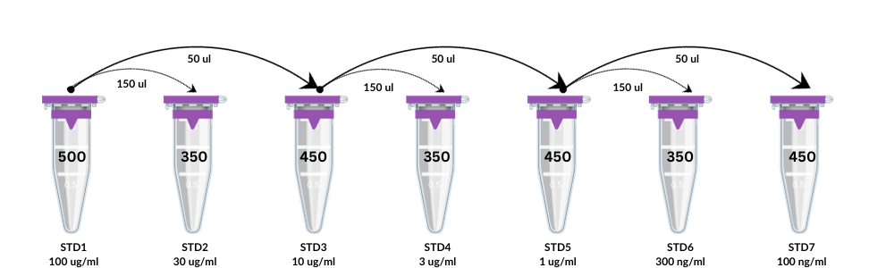
Prepare 500 ml of standard 1 in assay diluent at a final concentration of 100 ug/ml, according to the starting concentration on the vial. Prepare six tubes with indicated levels of assay diluent as shown in the table below. Transfer 150 ul of standard 1 into standard 2 and 50 ul of standard 1 in standard 3, mix thoroughly. Transfer 150 ul of standard 3 into standard 4 and 50 ul of standard 3 into standard 5, mix thoroughly. Transfer 150 ul of standard 5 into standard 6 and 50 ul of standard 5 into standard 7, mix thoroughly. Use assay diluent as a blank.
| Standard No. | Conc. (ug/ml) | Assay diluent (ul) | Standard (ul) | Final vol (ul) |
| Std-1 | 100 | 500 | 0 | 300 |
| Std-2 | 30 | 350 | 150 of S-1 | 500 |
| Std-3 | 10 | 450 | 50 of S-1 | 300 |
| Std-4 | 3 | 350 | 150 of S-3 | 500 |
| Std-5 | 1 | 450 | 50 of S-3 | 300 |
| Std-6 | 0.3 | 350 | 150 of S-3 | 500 |
| Std-7 | 0.1 | 450 | 50 of S-5 | 500 |
Sample collection and handling
Ideally, blood collection should be performed using standard venipuncture technique with vacutainers. Prior to testing, clots and all solid material should be removed by centrifugation. This assay was validated with plasma treated with K2 EDTA as an anti-coagulant. Conditioned media should be filtered using 1 uM filter plates prior to testing, to remove cell debris.
Avoid multiple freeze-thaw cycles as it may reduce the signal.
Sample dilution
For plasma samples, the recommended starting dilution is 4x. For purified conditioned media, the dilution varies depending on the cell type. Use assay diluent to dilute samples and always allow 10% extra to account for pipetting errors.
Detection antibody
In a benchtop centrifuge, briefly spin down detection stock, vortex 10 seconds. For one plate, add 26 ul from the detection stock to 5174 ul assay diluent, to reach a final volume of 5200 ul. Vortex the mixture and keep it on ice until used.
Streptavidin-phycoerythrin (SAPE)
The reagent is supplied at 10x concentration.
Assay diluent
The reagent is supplied at the working concentration.
Plate washing
Handheld magnet:
- Place the assay plate on the magnetic separator plate. Allow 2 min for the beads to separate from the solution onto the magnet.
- Carefully holding the assay plate on the magnetic separator so that the magnetic field is not disrupted, invert over an appropriate waste receptacle and gently shake out fluid, then blot out the excess on absorbent pads.
- With the plate still on the magnetic separator, add 200 ul wash buffer (PBS-T) to each well and cover the plate with an opaque lid. Allow 2 mins for separation.
- Repeat the wash steps as recommended in the assay protocol.
Magnetic plate washer:
Please refer to the manufacturer’s recommendations for programming instructions.
Equipment settings
This assay is compatible with Luminex200, HTS, Flexmap 3D, and MagPix with xPonent software; and xMap Intelliflex with Intelliflex software.
Please note that each instrument must be calibrated, and performance verified with the appropriate kits supplied by Luminex. In addition, the probe height must be adjusted to the plate provided in the kit for optimal bead count.
The specifications provided are for the instruments and software listed above:
- Events: 100 per each capture bead type (region)
- Sample size: 75 ul
- Gate settings: 5000-20000
- Bead type: magplex
- Reporter gain: default
- Time out: 60 seconds
| Reagent | Catalog No. | Region |
| CD9 | C-01-tspn | 25 |
| CD63 | C-02-tspn | 57 |
| CD81 | C-03-tspn | 48 |
| Control | C-00-tspn | 20 |
Assay protocol
Before beginning, please read all instructions and create a reference map of the 96-well plate with standards (low EV content), standards (high EV content), blanks, and samples. Follow the reagent preparation section and plate washing section for more details.
- Add 200 ul PBS to each well of the assay plate. Invert the plate over a sink, shake and blot out excess on paper towels.
- Prepare required amount of capture antibody mix in assay diluent. Incubate for 1 hour on a tumbler (12 rpm) at room temperature (20-25°C) before adding to samples.
- While incubating, prepare samples and standards, to be tested in duplicates. Allow 10% extra volume for pipetting errors.
- Vortex and sonicate capture antibody mix, add to assay plate (50 ul/well).
- Add samples and standards to assay plate (50 ul/well), according to plate map.
- Add assay diluent to background wells.
- Seal with black film and incubate overnight (16-20 hours) at 4°C on an oscillating shaker (500 rpm).
- Prepare wash buffer (1x PBS-T).
- Centrifuge the assay plate for 2 minutes at 1000rpm.
- Prepare the required amount of detection antibody mix in assay diluent.
- Wash the assay plate 3 times on a magnetic separator with 200ul wash buffer, as described in the plate washing section.
- Add detection antibody mix to assay plate (50 ul/well).
- Seal with black film and incubate for 2 hours at room temperature (20-25°C) on an oscillating shaker (500 rpm).
- Wash the assay plate 3 times on a magnetic separator with 200ul wash buffer, as described in the plate washing section.
- Add streptavidin-phycoerythrin to assay plate (50 ul/well).
- Seal with black film and incubate for 20 minutes at room temperature (20-25°C) on an oscillating shaker (500 rpm).
- Prepare 1x sheath fluid.
- Wash the assay plate 3 times on a magnetic separator with 200ul wash buffer, as described in the plate washing section.
- Add sheath fluid to assay plate (100 ul/well). Seal with black film and shake for 5 minutes at room temperature (20-25°C) on an oscillating shaker (500 rpm).
- Read the plate on Luminex200, HTS, Flexmap 3D, and MagPix with xPonent software; and xMap Intelliflex with Intelliflex software.