



How Do You Add a Brain-Specific Biomarker to a Trial Panel?
Adding a Tissue-of-Origin Biomarker to an Existing Trial Panel Brain-Specific Biomarker
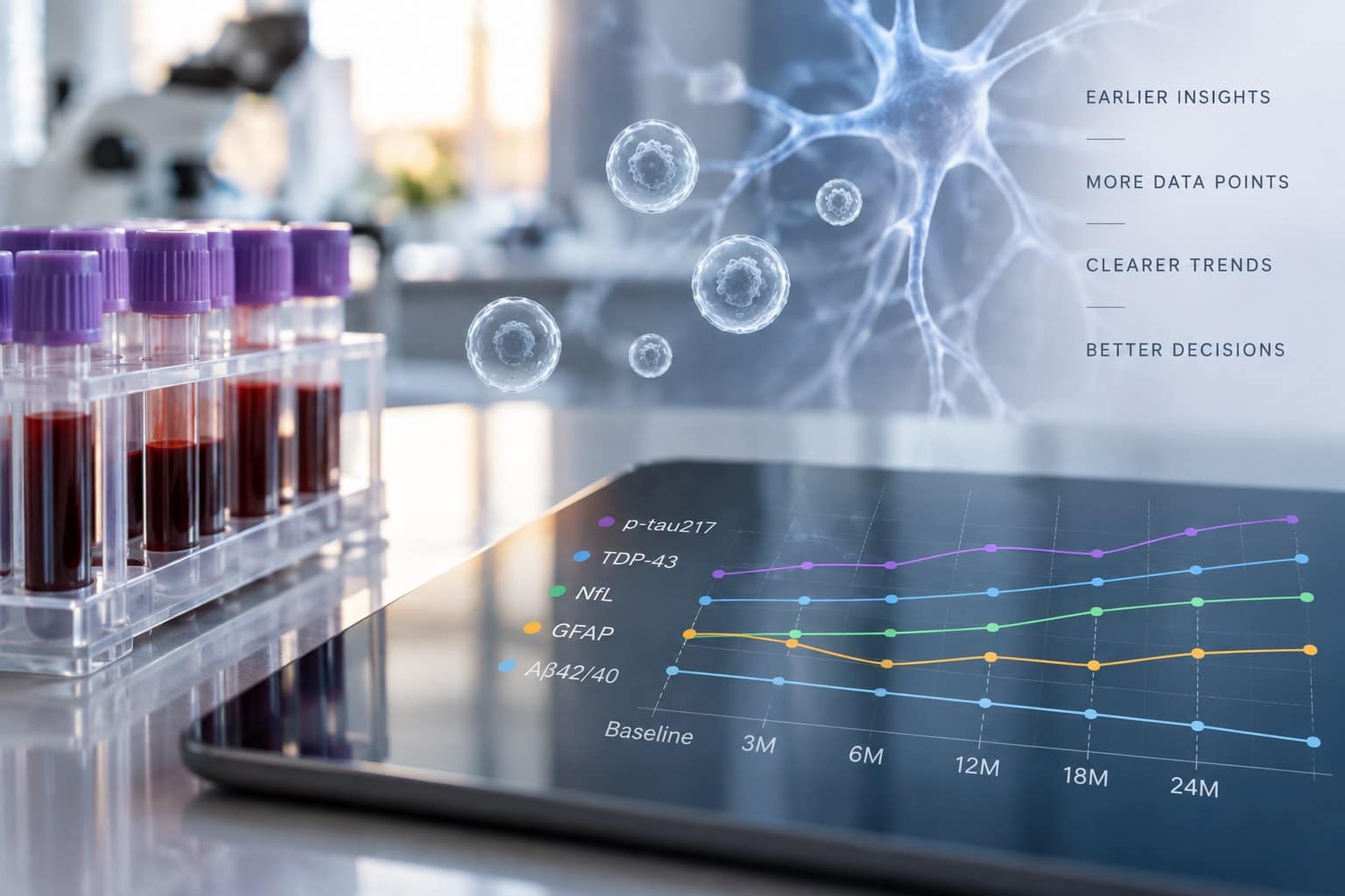
A Phase II readout comes back. Plasma neurofilament light moved in the treated arm. So did p-tau. The effect is nominally significant, the assay CVs are clean, and the central lab did nothing wrong. Then someone in the biomarker review asks the question that stops the meeting: how much of that movement came from the brain? Nobody can answer. The panel was never designed to answer it. The signal is real, but its origin is undetermined — and an undetermined origin cannot support a claim about a CNS mechanism.
Key Takeaways
- Brain specificity is a property of sample preparation, not of the analyte. Most proteins on a CNS panel are expressed outside the nervous system, so a plasma concentration is a sum across tissues.
- The three main trial decisions tolerate mixed-origin signal differently. Enrichment is often survivable. Target engagement usually is not.
- A tissue-of-origin readout is additive. It sits alongside your existing panel rather than replacing it, and the two are most informative when read together.
- Most of the operational burden is pre-analytical, and most of it can be written into the lab manual at study start rather than added by amendment.
- The interpretive gain is a denominator, not just another number: brain-specific signal expressed against total plasma signal separates CNS change from systemic change.
What makes a blood biomarker brain-specific?
A blood biomarker is brain-specific when the material being measured has been separated by tissue of origin before the analyte is quantified — not when the analyte itself is unique to neurons. Because most CNS-relevant proteins are also expressed peripherally, specificity comes from isolating brain-derived material from the plasma first, then measuring the target within that fraction.
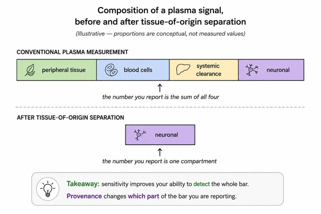
This distinction matters because the field often uses “specific” to mean “sensitive.” They are unrelated properties. An assay can detect single-digit femtomolar concentrations of a target and still be reporting a number in which brain-derived molecules are a minority contributor. Lowering the limit of detection improves your ability to see a signal. It does nothing to tell you where the signal came from.
The confound is compositional, not analytical
Consider three markers that appear on nearly every CNS trial panel.
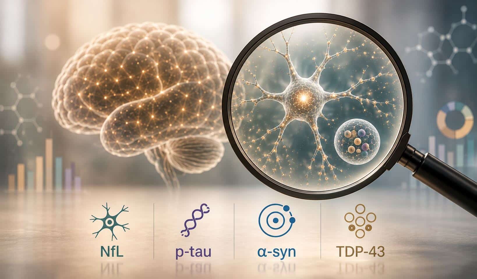
Neurofilament light chain. NfL is neuron-restricted, which is why it is often described as brain-specific biomarker. But neurofilaments are structural components of both central and peripheral axons. In a cross-sectional study of 75 patients with Charcot-Marie-Tooth disease and 67 matched controls, plasma NfL was roughly 78% higher in the patient group despite the pathology being confined to peripheral nerve [1]. A trial population with diabetic neuropathy, chemotherapy exposure, or comorbid peripheral nerve disease carries this contribution silently.
Alpha-synuclein. The compositional problem here is severe. The overwhelming majority of α-synuclein in whole blood is contained in erythrocytes, with plasma holding a small fraction of the total [2]. Variable hemolysis during collection or processing releases erythrocytic α-synuclein into the plasma compartment, where it can exceed the neuronal contribution by orders of magnitude [3]. The measurement is dominated by a pre-analytical variable rather than by biology.
Phosphorylated tau and TDP-43. Both are subject to systemic modifiers. Plasma p-tau217 concentrations rise with declining kidney function, and in a Mayo Clinic cohort analysis the effect was large enough to shift classification of amyloid status in individuals with CKD stage 3b or worse [4]. Broader comorbidity analyses show similar patterns across amyloid and neurodegeneration markers [5], and renal function affects NfL and p-tau181 in community cohorts as well [6]. TDP-43 presents the mirror image of the α-synuclein problem. Rather than being concentrated in one blood compartment, it is expressed broadly across tissue types, so a conventional plasma measurement carries a large and variable non-neuronal background — which is a substantial part of why TDP-43 has remained difficult to measure as a biofluid biomarker despite being the defining pathology in the great majority of ALS cases and a large share of FTD [12].
None of these are assay failures. Every one of them is a question about what is in the tube. And the question is not answerable after the fact: once a mixed-origin sample has been measured, there is no analytical or statistical operation that recovers the contribution attributable to the brain. Provenance is determined at the bench, before quantification, or it is not determined at all.
Figure 1. Composition of a plasma signal, before and after tissue-of-origin separation (Illustrative — proportions are conceptual, not measured values)
Three trial decisions that change with a brain-specific readout
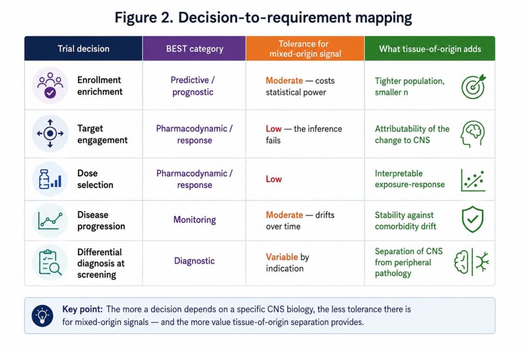
Panel design should follow from the decision the biomarker has to support, not from marker identity. The regulatory vocabulary here is useful: the FDA-NIH BEST resource separates diagnostic, prognostic, predictive, monitoring, pharmacodynamic/response, susceptibility, and safety biomarkers, and the evidentiary requirements differ by category [7,8]. A mixed-origin marker performs acceptably in some of these roles and poorly in others.
Enrichment and stratification
Enrichment asks a population-level question: does this patient belong in the trial? Here, a mixed-origin marker is often survivable. If elevated plasma NfL correlates with the disease state well enough to concentrate progressors in the enrolled population, the enrichment works even if some of the signal is peripheral. The cost of the confound is statistical dilution rather than a false conclusion — you enroll a somewhat noisier population and pay for it in sample size.
Tissue-of-origin separation still helps, particularly in populations with high comorbidity burden or in trials enrolling on the basis of a marker with a large systemic component. But this is the decision most tolerant of mixed origin.
Target engagement and pharmacodynamics
This is where mixed-origin readouts break first, and break hardest.
A pharmacodynamic biomarker is meant to demonstrate that a biological response occurred following exposure to the product [7]. The inference is causal and specific: the drug reached the compartment, engaged the target, and changed something measurable. A plasma concentration that sums contributions across tissues cannot support that inference. If the number moves, the movement is compatible with CNS target engagement, with a peripheral effect of the same drug, with a change in renal clearance, or with a shift in sample handling across visits.
For a CNS program using a plasma readout as proof of mechanism, this is not a precision problem. It is an interpretability problem, and no amount of analytical improvement resolves it. The measurement has to be attributable to the brain before a change in it can be attributed to CNS engagement.
The consequence shows up in dose selection. If the exposure-response relationship is built on a readout that sums CNS and peripheral contributions, the curve you fit is a composite of two response surfaces that may have different shapes and different EC50s. Choosing a Phase III dose from that curve is a coin flip dressed as an analysis. This is the specific scenario in which the cost of adding a second tier is trivially small relative to the cost of being wrong.
Progression and monitoring
Longitudinal monitoring sits between the two. Within-subject change partially controls for stable peripheral contributions — a patient’s erythrocyte mass and baseline peripheral nerve status do not swing wildly between visits. But comorbidities progress, kidney function declines, and hemolysis varies by draw. Over a two-year study, the peripheral contribution is not a constant to be subtracted.
Figure 2. Decision-to-requirement mapping
What you add, and what stays
The common misreading of this argument is that a brain-specific readout replaces the plasma panel. It does not, and a program that swaps rather than adds gives up information it already paid for.
Extracellular vesicles provide the practical route to tissue-of-origin separation in blood. EVs are membrane-bound particles released by essentially all cell types, carrying cargo that reflects the state of the originating cell, and their surface composition carries information about that cell of origin [9]. Immunocapture against neuronal surface epitopes therefore allows a population enriched for neuron-derived vesicles to be separated from bulk plasma, and the target protein quantified within that population. Work in this area has demonstrated that plasma EVs carry measurable TDP-43 and full-length tau, supporting discrimination among FTD, ALS, and progressive supranuclear palsy in multi-cohort analyses [10]. NeuroDex’s ExoSORT™ platform performs this enrichment step in an automated plate-based workflow ahead of downstream immunoassay.
The field is not uniformly settled, and it should not be presented as such. A systematic review and diagnostic meta-analysis of CNS-enriched EV biomarkers in parkinsonian disorders found considerable methodological heterogeneity across published studies and cautioned against over-interpreting pooled performance estimates [11]. That critique is a reason to demand isolation-specificity data and standardized reporting from any provider, not a reason to keep measuring bulk plasma.
Practically, the panel architecture becomes two tiers:
- Tier 1 (retained): your existing plasma panel, run by the central lab as currently specified. This continues to provide the systemic view, comparability to published cohorts, and safety-adjacent monitoring.
- Tier 2 (added): the tissue-of-origin readout, run on a parallel aliquot, reporting the same or overlapping analytes within the neuron-derived fraction.
The two tiers answer different questions. Tier 1 tells you the total burden. Tier 2 tells you how much of it is brain.
Sample handling and operational requirements
Most of the effort in adding a second tier is pre-analytical, and most of it is manageable if specified at study start rather than retrofitted.
The requirements that typically differ from routine chemistry:
- Anticoagulant and tube type, specified consistently across sites
- Time from draw to centrifugation, with a defined ceiling, since delay increases cellular contribution
- Centrifugation protocol, generally a defined two-step spin to remove cells and large debris
- Aliquot volume and count, sized so the second tier does not compete with Tier 1 for sample
- Freeze-thaw ceiling and storage temperature, documented per aliquot
- Hemolysis assessment, which matters more for some analytes than others and should be recorded rather than assumed
None of this is exotic. It is the pre-analytical discipline that MISEV2023 codified for EV studies generally, and it maps onto standard central-lab practice with a modest increase in specification detail [9]. The consensus document is worth putting in front of whoever writes your lab manual.
The operational decision worth making early is whether the second tier runs in batch at study end or at interim timepoints. Batched terminal analysis is cheaper and reduces run-to-run variability. Interim analysis gives you a chance to act on the readout, but commits you to bridging across analytical batches. For a target-engagement application, interim readouts are usually worth the added complexity, because a pharmacodynamic signal that arrives after database lock cannot inform dose selection.
Reading a brain-specific number alongside your existing panel
The interpretive gain is not simply an additional column. It is a ratio.
When the same analyte is quantified both in bulk plasma and in the neuron-derived fraction, the relationship between the two becomes the informative quantity. Three patterns are worth pre-specifying in the statistical analysis plan:
Both move together. Total plasma signal and brain-derived signal change in the same direction and similar proportion. The systemic change is likely CNS-driven, and the conventional marker is behaving as a reasonable proxy in this population.
Brain-derived moves, total does not. A CNS effect is present but diluted below detection in the mixed-origin measurement. This is the pattern that argues most strongly for the second tier, and the one most often missed by plasma-only panels in early-phase work.
Total moves, brain-derived does not. Something systemic changed — renal function, a peripheral drug effect, a shift in sample handling. Without the second tier, this would likely have been reported as a CNS finding.
That third pattern is the one that should motivate the addition. It is the false-positive case, and it is invisible to a panel that cannot resolve origin.
Regulatory acceptance of tissue-of-origin readouts is still developing. No neuron-derived EV measurement currently holds formal biomarker qualification, and any program treating one as a primary endpoint should be planning early regulatory engagement rather than assuming precedent. But the direction of travel in the field is clear: as blood-based CNS measurement moves from feasibility to decision-making, the questions being asked of it have shifted from “can we detect this” to “can we attribute it.” Sensitivity answered the first question. Provenance is what answers the second.
References
[1] Sandelius Å, Zetterberg H, Blennow K, Adiutori R, Malaspina A, Laura M, Reilly MM, Rossor AM. Plasma neurofilament light chain concentration in the inherited peripheral neuropathies. Neurology. 2018;90(6):e518–e524. https://doi.org/10.1212/WNL.0000000000004932
[2] Tian C, Liu G, Gao L, Soltys D, Pan C, Stewart T, Shi M, Xie Z, Liu N, Feng T, Zhang J. Erythrocytic α-synuclein as a potential biomarker for Parkinson’s disease. Translational Neurodegeneration. 2019;8:15. https://doi.org/10.1186/s40035-019-0155-y
[3] Yu Z, Liu G, Zheng Y, Huang G, Feng T. Erythrocytic alpha-synuclein as potential biomarker for the differentiation between essential tremor and Parkinson’s disease. Frontiers in Neurology. 2023;14:1173074. https://doi.org/10.3389/fneur.2023.1173074
[4] Bornhorst JA, Lundgreen CS, Weigand SD, Figdore DJ, Wiste H, Griswold M, Vemuri P, Graff-Radford J, Knopman DS, Cogswell P, Jack CR, Petersen RC, Algeciras-Schimnich A. Quantitative assessment of the effect of chronic kidney disease on plasma p-tau217 concentrations. Neurology. 2025;104(3):e210287. https://doi.org/10.1212/WNL.0000000000210287
[5] Syrjanen JA, Campbell MR, Algeciras-Schimnich A, et al. Associations of amyloid and neurodegeneration plasma biomarkers with comorbidities. Alzheimer’s & Dementia. 2022;18(6):1128–1140. https://doi.org/10.1002/alz.12466
[6] Wu J, Xiao Z, Wang M, Wu W, Ma X, Liang X, Zheng L, Ding S, Luo J, Cao Y, Hong Z, Chen J, Zhao Q, Ding D. The impact of kidney function on plasma neurofilament light and phospho-tau 181 in a community-based cohort: the Shanghai Aging Study. Alzheimer’s Research & Therapy. 2024;16. https://doi.org/10.1186/s13195-024-01401-2
[7] FDA-NIH Biomarker Working Group. BEST (Biomarkers, EndpointS, and other Tools) Resource. Silver Spring, MD: Food and Drug Administration; 2016–. https://www.ncbi.nlm.nih.gov/books/NBK326791/
[8] Cagney DN, Sul J, Huang RY, Ligon KL, Wen PY, Alexander BM. The FDA NIH Biomarkers, EndpointS, and other Tools (BEST) resource in neuro-oncology. Neuro-Oncology. 2018;20(9):1162–1172. https://doi.org/10.1093/neuonc/nox242
[9] Welsh JA, Goberdhan DCI, O’Driscoll L, Buzás EI, Blenkiron C, Bussolati B, et al. Minimal information for studies of extracellular vesicles (MISEV2023): from basic to advanced approaches. Journal of Extracellular Vesicles. 2024;13(2):e12404. https://doi.org/10.1002/jev2.12404
[10] Chatterjee M, Özdemir S, Fritz C, Möbius W, Kleineidam L, Mandelkow E, et al. Plasma extracellular vesicle tau and TDP-43 as diagnostic biomarkers in FTD and ALS. Nature Medicine. 2024;30(6):1771–1783. https://doi.org/10.1038/s41591-024-02937-4
[11] Taha HB, Bogoniewski A. Analysis of biomarkers in speculative CNS-enriched extracellular vesicles for parkinsonian disorders: a comprehensive systematic review and diagnostic meta-analysis. Journal of Neurology. 2024;271(4):1680–1706. https://doi.org/10.1007/s00415-023-12093-3
[12] Dellar ER, Nikel L, Fowler S, Vahsen BF, Dafinca R, Feneberg E, Talbot K, Turner MR, Thompson AG. Extracellular vesicles in TDP-43 proteinopathies: pathogenesis and biomarker potential. Molecular Neurodegeneration. 2025;20. https://doi.org/10.1186/s13024-025-00859-4
Brain-specific biomarker
Leave a Reply