




Closing the Diagnostic Gap in Alzheimer’s: How Neuron-Derived Biomarkers Are Changing What’s Measurable
The last several years produced the first validated blood tests for Alzheimer’s pathology — a genuine milestone for a field that spent decades relying on lumbar punctures and PET scans to confirm what’s happening in the brain. But a validated blood test for amyloid or tau is not the same as a blood test that tells a clinical trial team which patient, carrying which combination of underlying pathology, is standing in front of them.
Key Takeaways
- Plasma p-tau217 immunoassays now reach diagnostic accuracies approaching CSF and PET for detecting amyloid pathology — a genuine shift in what’s clinically accessible.
- Most people with clinically diagnosed Alzheimer’s carry co-existing pathology — TDP-43, alpha-synuclein, or both — that today’s blood tests were not built to resolve.
- The unresolved problem isn’t assay sensitivity. It’s that most blood biomarkers can’t separate neuron-derived signal from the much larger pool of peripheral, non-brain protein circulating in plasma.
- Neuron-derived extracellular vesicle (NDEV) isolation is one approach to closing that provenance gap — it’s the mechanism behind NeuroDex’s ExoSORT™ platform.
Why aren’t Alzheimer’s blood biomarkers enough to diagnose the disease precisely?
A positive plasma biomarker like p-tau217 confirms amyloid pathology is present, but it can’t tell a trial team whether TDP-43 or alpha-synuclein co-pathology — present in a large share of diagnosed Alzheimer’s cases — is also driving a patient’s decline, because most Alzheimer’s blood biomarkers can’t separate neuron-derived signal from non-neuronal plasma sources.
The Decade Alzheimer’s Blood Biomarkers Changed Diagnosis
It’s worth being direct about how far the field has come, because the progress is real. Plasma phosphorylated tau at threonine 217 (p-tau217) now detects amyloid pathology with diagnostic accuracies in the 0.93–0.96 AUC range in secondary care settings — performance that rivals cerebrospinal fluid testing and positron emission tomography, at a fraction of the cost and invasiveness [1]. That finding has been corroborated in other large validation cohorts assessing plasma p-tau217 immunoassays against confirmed Alzheimer’s pathology [2], and head-to-head work distinguishing Alzheimer’s from other neurodegenerative disorders using plasma p-tau217 alone has shown similarly strong discrimination [3]. These assays are now recommended in current diagnostic and research frameworks for the disease, and their arrival has coincided with a wave of amyloid-targeting therapies that depend on knowing, early and reliably, which patients actually have amyloid pathology.
The real-world data behind that headline number is worth sitting with. In a validation study spanning nearly 1,800 participants across five European cohorts, plasma p-tau217 reached accuracies of 89 to 91 percent in secondary care and 85 percent in primary care settings, using a fully automated commercial immunoassay platform [1]. A two-cutoff approach — flagging results as positive, negative, or indeterminate rather than forcing a binary call — pushed accuracy up to 92 to 94 percent, at the cost of excluding 12 to 17 percent of samples as intermediate [1]. That’s the kind of tradeoff a trial statistician recognizes immediately: better accuracy on the samples you keep, in exchange for a meaningful share of screened patients you can’t act on with confidence. Accuracy was also lower — 83 percent — in participants aged 80 and older, precisely the age band that makes up a large share of the Alzheimer’s population [1]. None of this undermines what p-tau217 has done for the field. It does mean the current generation of whole-plasma assays, however well validated, still leaves a nontrivial share of real-world patients in ambiguous territory.
For a translational scientist evaluating a CNS asset, this matters enormously. A decade ago, confirming amyloid or tau status meant a lumbar puncture or a PET scan — both expensive, both logistically difficult to repeat at scale across multi-site trials. Today, a blood draw can do a version of that job. That’s not a small thing.
What Blood Biomarkers Still Can’t Tell You
Here’s the part that gets less attention: a positive amyloid or tau signal tells you a patient has Alzheimer’s-type pathology. It does not tell you whether that’s the whole story. Autopsy-based community studies have found that Alzheimer’s-related pathology overlaps with other proteinopathies in a substantial share of cases — reported mixed-pathology prevalence ranges from roughly 10% to 74% depending on how mixed pathology is defined and which proteins are screened for [4]. TDP-43 co-pathology alone shows up in a meaningful fraction of clinically diagnosed Alzheimer’s cases, and a recent real-world cohort study using cerebrospinal fluid seed amplification assays found that roughly a third of biomarker-confirmed early Alzheimer’s patients tested positive for alpha-synuclein co-pathology, despite having no clinical signs of a Lewy body disorder [5].
That matters for reasons beyond diagnostic accuracy. Co-pathology has been linked to faster cognitive decline and different treatment response patterns than “pure” Alzheimer’s pathology [5]. A trial that stratifies patients on amyloid or tau status alone, without visibility into what else might be driving a given patient’s decline, is working with an incomplete picture — and an incomplete picture at the patient-selection stage is exactly the kind of thing that erodes statistical power and confounds endpoint interpretation downstream. It’s also worth noting that co-pathology detection often depends on clinical suspicion in the first place: alpha-synuclein co-pathology in the cohort above was identified in patients with no clinical signs pointing toward a synucleinopathy, which means routine screening, not just symptom-triggered testing, is what surfaces it [5].
There’s a pharmacodynamic version of this same problem, and it matters just as much to a translational team as the diagnostic one. A trial testing a mechanism-specific therapeutic — an anti-tau antibody, an alpha-synuclein-targeting molecule, an anti-inflammatory agent aimed at neuroinflammation — needs a way to show that the drug is doing what it’s supposed to do in the tissue it’s supposed to do it in. If the blood-based readout for that mechanism is diluted by protein from liver, muscle, or circulating immune cells that have nothing to do with the drug’s target, a real pharmacodynamic effect can be masked by noise that has nothing to do with efficacy. That’s a different failure mode than a diagnostic false negative, but it traces back to the identical root cause: a biomarker that can’t confirm where its signal came from can’t cleanly separate target engagement from background.
Why Peripheral Signal Is the Real Barrier
The deeper issue is one of origin, not detection. Total tau measured in plasma correlates weakly with total tau measured in cerebrospinal fluid — a disconnect that puzzled the field for years, until research demonstrated that most circulating total-tau in blood originates from peripheral, non-brain tissue rather than the brain itself [6]. A team that generated an antibody selective for brain-derived tau found that this brain-specific signal correlated strongly with CSF measures (rho = 0.85) where total-tau in plasma did not (rho = 0.23), and that the brain-derived signal — unlike standard neurodegeneration markers — could distinguish Alzheimer’s from other neurodegenerative diseases regardless of co-existing pathology [6].
The scale of that specificity gain is worth spelling out. In the same study, brain-derived tau distinguished autopsy-confirmed Alzheimer’s disease from other neurodegenerative diseases with an area under the curve of 86.4 percent — while neurofilament light, one of the most widely used blood-based neurodegeneration markers, performed at 54.3 percent, close to chance [6]. Critically, the brain-derived signal’s diagnostic performance held regardless of whether a patient also carried other, concomitant pathology [6]. That last point is the one that matters most for a stratification problem: a biomarker’s usefulness shouldn’t degrade the moment a patient’s case gets biologically messy, because biologically messy is the norm in this disease, not the exception.
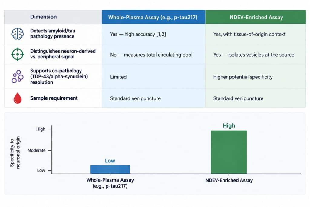
That finding reframes the whole problem. Even a validated, clinically useful biomarker like p-tau217 is being measured against a background of plasma that contains protein from liver, muscle, immune cells, and dozens of other non-neuronal sources. The assay can be exquisitely sensitive and still be diagnostically limited, because sensitivity to a protein isn’t the same as specificity to where that protein came from. This is the provenance problem behind plasma p-tau specificity: the binding constraint on precision Alzheimer’s diagnostics isn’t how well an assay can detect a molecule in plasma. It’s whether the plasma sample itself carries a signal that’s traceable back to the brain.
Isolating Signal at the Source: An NDEV-Based Approach
Neuron-derived extracellular vesicles (NDEVs) — small membrane-bound vesicles shed by neurons that cross the blood-brain barrier into peripheral circulation — offer one way around the provenance problem. Because they carry cargo from the neurons that released them, they function as a molecular snapshot of neuronal state, sampled non-invasively from blood. Early work using this approach identified elevated levels of pathogenic tau and amyloid species in neurally-derived blood exosomes years before clinical Alzheimer’s diagnosis, in some cases up to a decade ahead of symptom onset [7].
The magnitude of that early signal is part of why the approach generated so much interest in the first place. In the foundational case-control study, levels of three pathogenic proteins — two phosphorylated tau species and amyloid-beta 1-42 — measured in neurally-derived blood exosomes correctly classified more than 96 percent of Alzheimer’s patients using discriminant modeling, and the same protein signature was elevated in cognitively normal individuals who went on to develop Alzheimer’s, in some cases a full decade before diagnosis [7]. That’s a striking result for a field that, at the time, had no blood-based option even remotely competitive with CSF or imaging.
That promise comes with a caveat the field has had to reckon with directly: not every method for isolating neuron-derived vesicles from plasma performs consistently. A widely used single-surface-marker immunocapture approach was shown in a 2021 validation study to co-elute predominantly with free, non-vesicle-associated protein rather than with established extracellular vesicle markers — a finding that called into question a meaningful share of prior NDEV literature that relied on that single marker for enrichment [8]. The lesson wasn’t that NDEV isolation doesn’t work; it’s that the isolation method itself has to be validated against orthogonal markers and rigorously tested for what it’s actually capturing, not just what it was presumed to capture.
Validating an isolation method against orthogonal markers isn’t a one-time checkbox — it’s an ongoing discipline, because the field’s understanding of which surface proteins reliably track vesicle membranes, rather than free, cleaved, or soluble forms, continues to evolve. A platform’s credibility rests less on the specific markers it targets and more on whether it can demonstrate, batch after batch, that what it’s pulling out of plasma is actually vesicle-bound and actually neuronal in origin — not just present in the sample at an appropriate concentration.
This is the mechanism-level thesis behind NeuroDex’s ExoSORT™ platform: isolating neuron-derived vesicles from plasma using a validated, multi-marker enrichment approach, designed specifically to address the reproducibility gaps that single-marker methods have run into. The goal isn’t just to detect a protein in blood — it’s to confirm, at the point of isolation, that the signal being measured actually originated in neuronal tissue, separating it from the much larger background of peripheral protein that limits whole-plasma assays. That distinction is what determines whether a biomarker can meaningfully stratify a patient who is amyloid-positive and TDP-43-positive from one who is amyloid-positive alone — a distinction whole-plasma testing, however sensitive, structurally cannot make on its own.
What a Cleaner Signal Could Change in Practice
Picture two trial applications side by side. The first is patient selection: a sponsor enrolling a Phase II cohort for an amyloid-targeting or tau-targeting therapy wants to exclude patients whose cognitive decline is substantially driven by undetected TDP-43 or alpha-synuclein pathology, because those patients are less likely to respond to a mechanism that only addresses one piece of a mixed picture — and their inclusion dilutes the treatment effect the trial is powered to detect. A biomarker that can flag neuron-derived signal specific to each of those pathologies, rather than a single composite readout of “neurodegeneration,” gives a sponsor a more defensible basis for that kind of enrichment.
The second is longitudinal monitoring: a trial tracking treatment response over 18 to 24 months needs a biomarker stable and specific enough that a real signal change isn’t lost in assay noise or peripheral variability from one blood draw to the next. Both applications point toward the same requirement — not more sensitivity, but more confidence that the signal being tracked actually originated where the biology of interest is happening. This is the same logic behind how blood-based CNS biomarkers are reshaping trial endpointsmore broadly: narrowing variance, removing likely non-responders before dosing, and enabling denser sampling than serial lumbar punctures could ever support.
What This Means for Trial Design Going Forward
The next phase of Alzheimer’s biomarker development isn’t about finding a more sensitive assay for the same peripheral signal. It’s about building diagnostic and monitoring tools that can resolve co-pathology, track pharmacodynamic response at the level of neuronal biology, and support patient stratification models that account for the biological heterogeneity autopsy studies have documented for over a decade. As more amyloid-targeting therapies reach approval and more combination and mechanism-of-action trials move into the clinic, the sponsors who can answer “which pathology is this patient’s decline actually driven by” will be better positioned to interpret their own trial data — and to explain a null result as a stratification problem rather than an efficacy one.
There’s also a longer regulatory horizon worth naming. Biomarker qualification pathways with FDA and EMA reward validated, reproducible blood-based measures that can serve as objective tools across sponsors, rather than one-off assays run differently trial by trial. A biomarker that can demonstrate tissue-of-origin specificity, not just analyte sensitivity, has a stronger case for that kind of qualification — regulators evaluating a biomarker for use across multiple sponsors and multiple therapeutic mechanisms need confidence that a given signal means the same thing in every trial that uses it.
The disease is finally measurable in blood in ways it simply wasn’t a decade ago. The next milestone isn’t measuring more of the same signal. It’s measuring more precisely, from a source the field can actually trust.
Figure 1. Whole-Plasma vs. Neuron-Derived Vesicle Biomarker Approaches
Illustrative comparison table — source: synthesized from cited literature, not a single study’s dataset.
References
- Palmqvist S, Warmenhoven N, Anastasi F, et al. Plasma phospho-tau217 for Alzheimer’s disease diagnosis in primary and secondary care using a fully automated platform. Nat Med. 2025;31(6):2036-2043. https://doi.org/10.1038/s41591-025-03622-w
- Ashton NJ, Brum WS, Di Molfetta G, et al. Diagnostic accuracy of a plasma phosphorylated tau 217 immunoassay for Alzheimer disease pathology. JAMA Neurol. 2024;81(3):255-263. https://doi.org/10.1001/jamaneurol.2023.5319
- Palmqvist S, Janelidze S, Quiroz YT, et al. Discriminative accuracy of plasma phospho-tau217 for Alzheimer disease vs other neurodegenerative disorders. JAMA.2020;324(8):772-781. https://doi.org/10.1001/jama.2020.12134
- Rahimi J, Kovacs GG. Prevalence of mixed pathologies in the aging brain. Alzheimers Res Ther. 2014;6(9):82. https://doi.org/10.1186/s13195-014-0082-1
- Shiner T, Nathan T, Levy MH, et al. Alpha-synuclein co-pathology in a real-world early Alzheimer’s disease cohort. Alzheimers Dement. 2026;22(3):e71240. https://doi.org/10.1002/alz.71240
- Gonzalez-Ortiz F, Turton M, Kac PR, et al. Brain-derived tau: a novel blood-based biomarker for Alzheimer’s disease-type neurodegeneration. Brain.2023;146(3):1152-1165. https://doi.org/10.1093/brain/awac407
- Fiandaca MS, Kapogiannis D, Mapstone M, et al. Identification of preclinical Alzheimer’s disease by a profile of pathogenic proteins in neurally-derived blood exosomes: a case-control study. Alzheimers Dement. 2015;11(6):600-607.e1. https://doi.org/10.1016/j.jalz.2014.06.008
- Norman M, Ter-Ovanesyan D, Trieu W, et al. L1CAM is not associated with extracellular vesicles in human cerebrospinal fluid or plasma. Nat Methods.2021;18(6):631-634. https://doi.org/10.1038/s41592-021-01174-8

Leave a Reply