Cell Based Assay Services for Accelerated Drug Discovery
Think of a new drug’s journey to the clinic. Before it ever gets near a human patient, it has to prove its worth. Cell based assay services act as a kind of biological dress rehearsal, using living cells to see how a new compound might actually behave inside the body. It’s our first, best glimpse into a drug’s potential for success or failure.
How Cell Based Assays Accelerate Drug Discovery

cell based assay services
In the high-stakes world of drug development, most candidates don’t make it to the finish line. A big reason for this expensive failure rate is that early lab tests often can’t predict what will happen in the complex, dynamic environment of the human body. This is exactly where cell based assays are changing the game.
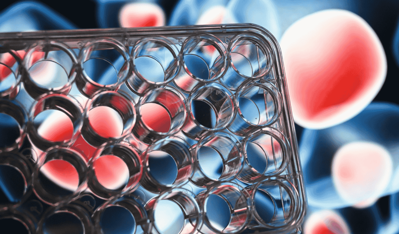
Unlike traditional biochemical tests, which look at molecules in isolation, cell-based assays put living cells to work as tiny, functional test subjects. This gives us a much richer, more meaningful picture of a drug’s real effects—from its intended therapeutic action to any unintended toxicity.
From Test Tubes to Living Systems
Imagine the difference between testing a car engine on a stand versus actually driving the car on a real road. A biochemical assay is like testing the engine alone; it tells you if the individual parts are working correctly.
A cell-based assay, on the other hand, is the road test. It shows you how that engine performs as part of the whole system, revealing subtle issues you’d completely miss otherwise.
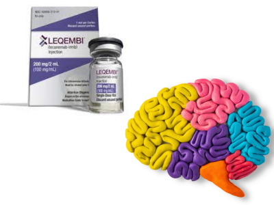
This shift is a massive deal, especially in complex fields like neurology. Brain diseases aren’t about one molecule misbehaving; they involve intricate networks of neurons, glia, and other cells all communicating with each other. A cell-based model can begin to mimic this environment, offering insights that are simply impossible to get from a reaction in a test tube.
Because of this, researchers can now make much smarter “go/no-go” decisions much earlier in the pipeline. It saves an incredible amount of time and money.
The industry has taken notice. The global cell-based assay market is on track to hit USD 32.02 billion by 2031, with a strong compound annual growth rate of 8.67%. This boom is driven by a clear demand for better tools to de-risk the notoriously expensive drug discovery process.
Core Applications of Cell Based Assay Services in R&D
Cell-based assays are not a one-trick pony. They’re a versatile platform used at nearly every stage of research and development to answer critical questions. These assays are the workhorses that help researchers decide which projects to push forward and which to shelve before they become costly dead ends.
The table below breaks down the key moments where these services provide the most value.
| Application Area | Key Objective | Typical Assay Used | Impact on Drug Development |
|---|---|---|---|
| Target Validation | Confirm a protein or pathway is truly linked to a disease. | CRISPR/siRNA knockdowns, reporter gene assays | Prevents investment in drugs aimed at the wrong biological target. |
| Lead Optimization | Screen thousands of compounds to find the most effective and safest. | High-content screening (HCS), cytotoxicity assays | Narrows the field to a few top candidates with the best therapeutic profile. |
| Safety & Toxicity | Identify harmful effects on cells early in the process. | Apoptosis assays, mitochondrial toxicity assays | Weeds out dangerous compounds long before they can reach human trials. |
By giving us a window into what’s happening at the cellular level, these assays allow research teams to fail faster and, more importantly, smarter. This focus on only the most promising candidates is how we get new therapies to patients who need them more quickly and efficiently.
Of course, generating all this biological data is only half the battle. To make sense of it, you need powerful analytical tools. That’s where the applications of bioinformatics in the pharmaceutical industry come in, turning massive datasets into actionable knowledge.
This synergy between wet-lab biology and data science is what powers modern drug discovery. When you combine these assays with broader strategies like those used in our biomarker discovery services, you get a complete, multi-layered view of a disease and how to treat it.
Choosing the Right Cell Model for Your Target
The single most important decision you’ll make in any cell-based assay is which cell model to use. It’s like casting the lead actor in a film—pick the wrong one, and the whole production can fall apart, no matter how good the script is. Getting this choice right ensures your data is relevant and predictive. Getting it wrong can send you down a dead end with misleading results that don’t translate to human biology.
The entire foundation of your assay rests on how well your chosen cells mimic the specific disease or biological process you’re studying. This decision is always a balancing act between capturing true biological complexity and maintaining practical scalability.
Starting with the Workhorses: Immortalized Cell Lines
For many assays, the most common starting point is an immortalized cell line. These are cells, like the famous HeLa or HEK293 lines, that have been engineered to divide indefinitely in a lab dish. They are the reliable workhorses of cell-based research.
Their main draws are consistency, ease of use, and scalability. Because they are so well-characterized and grow so quickly, they are perfect for high-throughput screening where you need to test thousands of compounds efficiently.
But their greatest strength is also their biggest weakness. After decades of continuous division, these cells have drifted significantly from their original genetic makeup. They often fail to accurately represent the intricate biology of a specific disease, especially in a complex field like neurology.
Creating a Miniature Ecosystem with Co-Culture Systems
To layer in more biological realism, researchers often turn to co-culture systems. Instead of growing just one type of cell, a co-culture combines multiple cell types in the same dish, creating a much better replica of a human tissue environment.
Think about it like this: trying to understand city traffic by watching only one car gives you some information, but you miss all the crucial interactions. A co-culture is like creating a miniature city grid, complete with different kinds of vehicles all interacting with one another.
In neurology research, this could mean growing neurons alongside astrocytes and microglia—the key support cells in the brain. This “miniature brain ecosystem” allows you to see how a drug affects not just the neurons, but the entire cellular neighborhood, revealing effects you would otherwise miss.
This approach delivers a much richer dataset by showing how cells communicate and influence each other. It’s a huge step up in biological relevance from a single cell line.
The Gold Standard: Patient-Derived iPSCs
For the highest level of human relevance, particularly in neurology, the gold standard is induced pluripotent stem cells (iPSCs). These are created by taking a skin or blood sample from a patient and “reprogramming” the cells back into a stem-cell-like state.
From that blank slate, scientists can then guide these iPSCs to become any cell type they need—in our world, that means specific types of brain cells like motor neurons or cortical neurons.
This is a game-changer for several reasons:
- Patient-Specific Genetics: The neurons carry the exact genetic makeup of the patient, including any mutations that cause their disease.
- Unprecedented Disease Modeling: You can create “disease-in-a-dish” models that faithfully replicate the cellular problems seen in diseases like Parkinson’s or ALS.
- Predictive Power: Testing a drug on these cells provides the most predictive data possible before moving into human trials. You are, in effect, testing the drug on a virtual version of the patient.
While iPSC-derived models are more complex and time-consuming to work with, the depth of insight they provide is invaluable. They bridge the gap between lab research and clinical reality, making them a cornerstone of modern cell based assay services for neurotherapeutics. These models are pushing the boundaries of what’s possible, much like the advancements seen in exploring 3D printed brain tissue for neuroscience insights.
Understanding Key Assay Readout Technologies
Once you’ve picked the right cell model, you face the next big question: how do you actually measure a drug’s effect? How can you tell what’s happening inside the cells? This is where assay readout technologies come into play. Think of them as sophisticated measurement tools that translate complex biological events into clear, actionable data.
Choosing the right readout is just as crucial as choosing the right cell model. These technologies are the high-tech eyes and ears of cell based assay services, letting researchers observe everything from a change in a cell’s shape to the electrical chatter between neurons. Each one offers a unique window into cellular function, providing different answers to specific biological questions.
This diagram shows how today’s increasingly complex cell models give us more biologically relevant platforms for these readouts.

The leap from simple cell lines to advanced iPSCs reflects the industry’s push toward models that better mimic human responses, making the data we get from these readouts far more translatable to the clinic.
High-Content Screening for Visual Data at Scale
Imagine automated microscopy running on a massive scale—that’s High-Content Screening (HCS). Instead of a scientist peering at one slide at a time, HCS systems use robotic microscopes and intelligent software to capture and analyze thousands of images from multi-well plates.
But this isn’t just about taking pretty pictures. HCS quantifies dozens of cellular features—the “content”—all at once. For a neurology study, that could mean measuring:
- The number and length of neuronal branches (neurites).
- Where a specific protein is located inside a cell, and how much of it is there.
- Subtle changes in the size or shape of the nucleus.
- The presence of toxic protein clumps, like those found in Alzheimer’s or Parkinson’s disease.
By turning images into rich, quantitative data, HCS lets researchers screen thousands of potential drugs to see which ones can clear those toxic proteins or encourage healthy neuron growth.
Electrophysiology Listening to Neuronal Conversations
If HCS is about seeing what cells look like, electrophysiology is about hearing what they’re saying. Neurons communicate using electrical signals, or action potentials, and Multi-electrode array (MEA) technology lets us “listen in” on these conversations in real-time.
An MEA plate has a grid of tiny electrodes at the bottom of each well. When you grow neurons on this surface, the electrodes can detect their electrical firing.
This allows scientists to assess the health and function of an entire neural network. A healthy network shows synchronized, rhythmic firing patterns, while a network damaged by disease or a toxic compound might fire erratically or fall silent.
In drug discovery, MEA is perfect for answering questions like, “Can this new drug restore normal electrical activity to diseased neurons?” or “Does this compound have unintended toxic effects on neural function?” It’s a direct functional readout that is absolutely critical for developing effective neurotherapeutics.
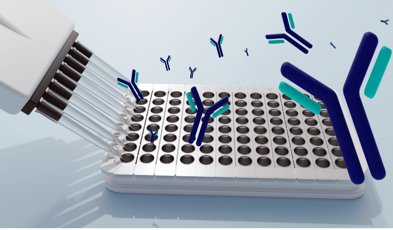
Reporter Gene Assays Cellular Light Switches
Another powerful tool is the reporter gene assay. This clever technique engineers cells to act like tiny light switches that turn on when a specific biological event happens. Researchers insert a “reporter gene”—often one that produces a light-emitting protein like luciferase—into the cell’s DNA.
This reporter gene is then tied to the activity of another gene you’re interested in. For example, to study an inflammation pathway, you could link the luciferase gene to an inflammatory response gene. When a drug activates that pathway, the cells produce luciferase and literally glow.
The intensity of the light directly corresponds to the activity of the pathway. This gives you a simple, highly sensitive, and easy-to-measure readout, making it ideal for screening huge numbers of compounds to find ones that turn a specific target on or off.
This kind of precision is a key reason the cell-based assay services market is poised for major growth. North America is currently leading the global market, with over 43% of the revenue, driven by strong R&D investment and a mature biopharma industry focused on advanced technologies like these.
Choosing the best readout depends entirely on the question you’re asking. To make it easier, here’s a quick comparison of the major technologies.
Comparison of Key Cell Assay Readout Technologies
| Technology | Measures | Best For | Throughput | Key Advantage |
|---|---|---|---|---|
| High-Content Screening (HCS) | Morphological changes (cell shape, protein location, neurite outgrowth) | Screening for phenotypic changes, visualizing compound effects at the subcellular level | High | Provides rich, multi-parametric visual data from a single experiment |
| Electrophysiology (MEA) | Neuronal electrical activity (firing rate, network synchrony, bursts) | Assessing neurotoxicity and efficacy, studying functional changes in neural networks | Medium | Gives a direct, real-time functional readout of neuronal communication |
| Reporter Gene Assays | Specific gene or pathway activation (e.g., via luciferase, GFP) | High-throughput screening for compounds that modulate a single, defined target | Very High | Extremely sensitive and easily quantifiable signal, ideal for large-scale screens |
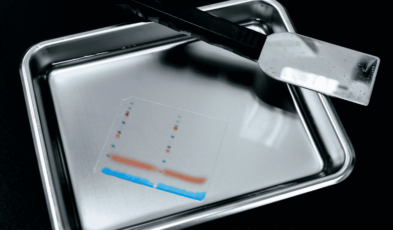
| ELISA / Immunoassays | Secreted proteins or intracellular protein levels (e.g., cytokines, biomarkers) | Quantifying specific protein production or release in response to a stimulus | High | Highly specific and quantitative, excellent for biomarker validation |
| Flow Cytometry | Cell populations, surface markers, cell cycle, apoptosis | Analyzing heterogeneous cell populations and rare cell events | High | Analyzes individual cells at high speed, providing population-level statistics |
Each of these technologies provides a different piece of the puzzle. HCS shows you what’s happening visually, MEA lets you listen to the functional output, and reporter assays give you a bright signal for a specific molecular event. Often, the most powerful insights come from combining several of these readouts. You can find more details in the full cell-based assays market report on these trends. It’s also helpful to see how ELISA kit production supports these sensitive assay readouts for protein-level quantification.
Building a Robust and Validated Assay Workflow
An idea for a cell-based assay is just the beginning. The real value in cell based assay services comes from turning that concept into a reliable, reproducible, and regulatory-ready tool. After all, an assay is only as good as its data, and generating trustworthy results demands a methodical, multi-stage workflow.
This process isn’t a single event but a journey through three critical phases: development, optimization, and validation. Each step builds on the last, systematically refining the assay until it performs with the precision of a finely tuned instrument. This is the bedrock of generating data that can confidently guide multi-million dollar drug development decisions.
Phase 1: Assay Development
The development phase is where we bring all the core components together for the first time. Think of it as the “proof-of-concept” stage. The primary goal is simple: show that the assay can actually measure the biological effect we’re interested in.
Key activities during this initial stage include:
- Selecting the right cell model and readout technology to answer the biological question.
- Establishing basic protocols for cell culture, compound treatment, and data acquisition.
- Identifying appropriate positive and negative controls to ensure the assay responds as expected.
- Confirming a detectable signal that changes when we introduce known modulators.
At this point, the assay works, but it isn’t yet precise. The signal might be weak or the variability high, which is where the next crucial phase comes in.
Phase 2: Tuning the Instrument Through Optimization
Optimization is where a functional assay becomes a high-performance one. It’s like tuning a sensitive radio to get the clearest possible signal, methodically eliminating static and noise. Here, the goal is to refine every variable to maximize the assay’s performance, sensitivity, and reproducibility.
This is an iterative process of systematically adjusting parameters to find the “sweet spot” for each one. We’ll often fine-tune things like reagent concentrations, incubation times, cell plating densities, and the timing of compound additions.
The objective is to achieve the largest possible assay window—the difference in signal between the positive and negative controls. A wide, clean window makes it easy to distinguish real biological effects from background noise, ensuring the data is unambiguous and reliable.
As part of a robust workflow, we can also integrate sustainable laboratory practices to optimize resource use and minimize environmental impact, reflecting both operational efficiency and corporate responsibility.
Phase 3: Validation for Regulatory Confidence
Validation is the final and most rigorous stage. This is where the assay is formally tested to prove it is fit for its intended purpose, especially if the data will be used in regulatory submissions. This process is governed by strict guidelines like Good Laboratory Practice (GLP).
GLP isn’t just a suggestion; it’s a quality system mandated by regulatory agencies like the FDA to ensure the uniformity, consistency, and integrity of non-clinical safety data. For IND-enabling studies, working with a GLP-compliant service provider is non-negotiable.
During validation, we establish and document key performance metrics:
- Accuracy: How close are the measurements to the true value?
- Precision: How consistent and repeatable are the results?
- Robustness: Can the assay handle small, deliberate variations in the method without failing?
- Specificity: Does it assess the target analyte without interference from other components?
A critical quality control metric we measure is the Z-factor (or Z-prime). This statistical value boils down the means and standard deviations of the positive and negative controls into a single number that reflects the assay’s quality. A Z-factor greater than 0.5 is widely considered the hallmark of an excellent, high-throughput screening assay, indicating a large separation between signals and very low data variability. This level of validation ensures that every data point is defensible, reliable, and ready for scrutiny.
Connecting Cell Assays to Clinical Biomarkers

Discoveries made in a lab dish are fascinating, but they only really matter if they predict what will happen in a human patient. This is the single biggest challenge in drug development—bridging that massive gap between a promising preclinical finding and actual clinical success. Modern cell-based assay services are now engineered to build this bridge from day one by aligning directly with a clear clinical biomarker strategy.
The game has changed. It’s no longer enough to find a compound that “works” in a cell model. The real goal is to design an assay that measures an effect you can also track in patients during a clinical trial. This forges a direct, translatable link between the lab bench and the bedside.
Creating a Seamless End-to-End Data Story
Let’s walk through an example. Imagine you’re developing a new drug for a neurodegenerative disease. Using a sophisticated iPSC-derived neuron model built from patient cells, your assay shows the drug successfully lowers the levels of a specific toxic protein inside those neurons. A decade ago, this is where the preclinical story might have ended, leaving a giant question mark: how do we measure that same effect in a living person?
Today, that story continues. The key is to connect the intracellular protein you measured in the assay to a biomarker that can be accessed with a simple blood draw. This is where advanced platforms that analyze neuron-derived exosomes (NDEs) become absolutely essential.
NDEs are tiny vesicles released by brain cells that travel into the bloodstream, carrying a molecular snapshot of what’s happening inside those cells. By isolating these NDEs from a blood sample, we can measure the exact same toxic protein you tracked in your initial cell-based assay.
This powerful connection establishes a seamless data pipeline. It allows you to follow a drug’s impact directly from the initial cell model all the way through human clinical trials, telling one continuous story.
The Power of Translatable Endpoints
Aligning preclinical assays with clinical biomarkers this way gives you an enormous advantage in de-risking your drug development program. It provides clear, evidence-based answers to the most critical questions you’ll face.
By integrating this strategy from the start, your cell-based assay services can help you:
- Verify Target Engagement: Confirm your drug is actually hitting its intended target in humans, not just in a lab model.
- Establish Dose-Response: Figure out the right dose needed to produce a measurable biological effect in patients.
- Stratify Patient Populations: Identify the patients who are most likely to respond to the treatment based on their specific biomarker profile.
- Monitor Drug Efficacy: Track the drug’s therapeutic effect over time with objective, quantitative data.
This approach transforms cell-based assays from isolated research tools into the foundational first step of a coherent, end-to-end clinical development plan. It ensures that the insights you gain in the lab aren’t lost in translation but instead serve as a reliable guide for making smarter, faster decisions in the clinic. The result is a dramatically improved probability of success, getting effective new therapies to patients who need them more efficiently.
How to Select the Right Service Partner
Choosing a partner for your cell-based assay services is a high-stakes decision. It’s a choice that can genuinely steer the direction of your entire drug development program. The right team becomes a true scientific collaborator, an extension of your own lab. The wrong one? They can hand you unreliable data that sends you down expensive, time-consuming dead ends.
This decision goes way beyond a simple price sheet. It’s about finding a group with the deep expertise and rock-solid quality systems needed to generate data you can build a program on.
A vendor simply offers a service. A partner, on the other hand, delivers a solution. The difference is their ability to grasp your scientific goals and proactively help you reach them.
Evaluating Technical Expertise and Experience
First things first: you have to vet their technical skills. Not all providers are set up to handle the complex, biologically meaningful models that today’s research demands, especially in a field as nuanced as neurology. A potential partner’s hands-on experience with specific cell types and disease models isn’t just a “nice-to-have”—it’s a dealbreaker.
When you’re talking to potential partners, get specific. Ask them pointed questions about their real-world experience:
- Complex Cell Models: Do they have proven, in-house expertise with advanced systems like iPSC-derived neurons, co-cultures, or 3D organoids? Don’t just take their word for it; ask to see case studies or data examples.
- Disease Area Track Record: Have they actually developed and validated assays in your specific therapeutic area, like neurodegeneration or neuroinflammation?
- Technological Capabilities: Can they support the exact readout technologies you need? Whether it’s high-content imaging or sensitive electrophysiology, make sure they have the right tools and the know-how to use them effectively.
A partner’s ability to work with patient-derived iPSCs, for instance, is a strong signal that they can handle the most translationally relevant—and often most challenging—models out there.
Assessing Quality Systems and Regulatory Compliance
Beyond technical chops, the single most critical factor is the integrity of their quality management system. If the data is going to support a regulatory filing, it absolutely must be generated under the strictest standards to ensure it’s reproducible, traceable, and unimpeachable.
A provider’s commitment to quality is non-negotiable. If the data from an assay will be part of an IND submission or other regulatory package, it must be generated under Good Laboratory Practice (GLP) standards. This is the baseline for creating defensible, high-integrity data that regulatory bodies like the FDA will accept.
Don’t be shy about auditing a potential partner’s quality systems. Look for clear documentation, rigorous SOPs, and a culture where quality control is just part of the DNA, embedded in every step of the workflow. Ask them how they qualify and validate their assays, and listen closely for metrics like the Z-factor, which is a key indicator of assay robustness.
Finally, think about the long-term picture. The best cell based assay services aren’t just one-off experiments; they’re designed with the end game in mind. An ideal partner will ask you how your preclinical assay data will eventually connect to your clinical biomarker strategy. That kind of foresight—the ability to help you build a seamless data story from the lab bench to the patient bedside—is what separates a mere vendor from a true scientific ally.
Your Questions, Answered
When you’re looking to outsource cell-based assays, a lot of practical questions come up—about how long things take, whether the data will mean anything in the clinic, and what all the regulatory jargon really means. Here are some of the most common questions we get from R&D teams and clinical trial sponsors, along with our straightforward answers.
What’s a Realistic Timeline for Developing a Custom Cell-Based Assay?
The timeline really depends on the complexity of what you’re trying to build.
For a relatively simple assay—say, using a standard, well-behaved cell line with a common readout—we can often get development and optimization done in just 4-6 weeks. This is perfect for those early-stage screening campaigns where speed is key.
But if you’re looking at something more sophisticated, like differentiating iPSCs into a specific type of neuron and then developing a novel high-content imaging analysis, you need a longer runway. A project like that can easily take 3-6 months to get fully developed, optimized, and validated under strict GLP standards. A good partner will always be upfront about this, giving you a detailed project plan with milestones that make sense for your specific target, cell model, and endpoints.
How Do You Make Sure Lab Data Actually Translates to Clinical Outcomes?
This is the most important question, and the answer comes down to two things: using clinically relevant cell models and building a smart biomarker strategy from the very beginning. The whole point is to forge a direct link between what you measure in a dish and what you can measure in a patient.
The best way to do this is to design your preclinical assay around an endpoint you can also track in the clinic. For example, if your iPSC-neuron assay shows your drug reduces a specific toxic protein, you should be able to measure that same exact protein in patients using a blood test that analyzes neuron-derived exosomes. This approach creates a seamless bridge, turning your preclinical data into a powerful predictor of how the drug will perform in people.
This alignment between preclinical assays and clinical biomarkers is the single most important factor in boosting the probability of clinical success. It transforms lab data from a simple observation into a predictive tool for human outcomes.
What’s the Real Difference Between GLP and Non-GLP Assays?
The distinction all comes down to the end goal. Non-GLP (Good Laboratory Practice) assays are built for speed and flexibility. They’re what you use for early-stage research, discovery work, and initial screening where you need to iterate quickly.
GLP assays, on the other hand, are performed under a rigorous, government-mandated quality system. This isn’t optional; it’s a strict framework ensuring every single data point is reproducible, traceable, and has the integrity needed for a regulatory submission. If your data is going to support an Investigational New Drug (IND) application or be sent to the FDA, it absolutely has to be GLP.
Can You Use Cell-Based Assays for Biologics and Cell Therapies?
Absolutely. In fact, they’re essential. Cell-based assays provide the biological context you need to figure out if these advanced therapies are actually working. You can assess their potency, understand their mechanism of action, and spot potential off-target effects.
A classic example is testing a CAR-T cell therapy. You can set up a co-culture assay that puts the CAR-T cells directly up against cancer cells. This gives you direct, functional data on the therapy’s tumor-killing ability long before it ever gets near a patient, making these assays indispensable for developing next-generation therapeutics.
At NeuroDex Inc, building these translational bridges is what we do. Our deep expertise in iPSC-derived neuronal models and neuron-derived exosome biomarkers means your preclinical data is designed for clinical success from day one. Learn how we can accelerate your neurotherapeutics program.
Leave a Reply