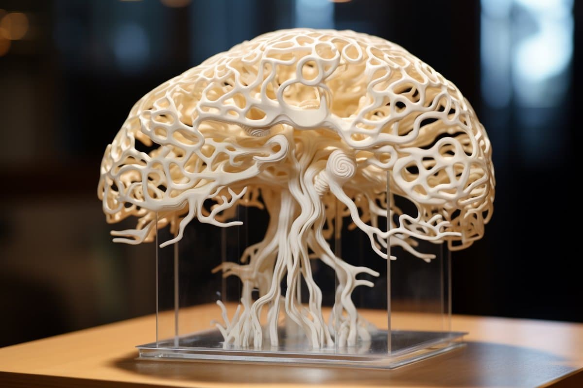
Revolutionary 3D-Printed Brain Tissue Offers New Insights into Neuroscience
Scientists have successfully created 3D-printed brain tissue that closely mimics the behavior of natural brain tissue. This development holds great promise for advancing research in neurological and neurodevelopment disorders.
The novel 3D-printing technique utilizes a horizontal layering method and a softer bio-ink, enabling the interconnection and formation of networks resembling those found in human brain structures.
Additionally, the 3D-printed brain tissue can be customized to mimic specific brain regions, allowing scientists to study the behavior and interactions of different brain regions. This level of control is not possible with traditional cell culture methods.
The researchers behind this breakthrough believe that their 3D-printed brain tissue could eventually replace animal models in neuroscience research. Animal models have limitations and may not accurately reflect human brain function and diseases.
By using 3D-printed brain tissue, scientists can more effectively study human-specific neurological processes and test potential treatments. This could lead to the development of targeted therapies for neurodevelopmental disorders and a better understanding of how certain drugs affect the brain.
Furthermore, this technology has the potential to revolutionize personalized medicine. By creating 3D-printed brain tissue from individual patients, doctors could test various treatment options and predict their efficacy before administering them to the patient.
While this breakthrough is still in its early stages, it represents a significant step forward in neuroscience research. The ability to create realistic and customizable 3D-printed brain tissue opens up new possibilities for studying the complexities of the human brain and developing innovative treatments for neurological disorders.
Key Facts:
- The 3D-printed brain tissue can form networks and communicate through neurotransmitters, similar to the interactions observed in the human brain.
- The new printing method provides accurate control over cell types and arrangements, exceeding the capabilities of traditional brain organoids.
- Many labs can use the technique without needing special equipment or culture methods, and it can significantly impact the study of various neurological conditions and treatments.
A team of University of Wisconsin–Madison scientists has developed the first 3D-printed brain tissue that can grow and function like typical brain tissue.
This achievement has significant implications for scientists studying the brain and working on treatments for various neurological and neurodevelopmental disorders, such as Alzheimer’s and Parkinson’s disease.
Su-Chun Zhang, professor of neuroscience and neurology at UW-Madison’s Waisman Center, suggests that this model can be highly beneficial in comprehending communication between the brain cells and different brain regions in humans.
“It could change the way we look at stem cell biology, neuroscience, and the pathogenesis of many neurological and psychiatric disorders.”
The researchers at the University of Wisconsin-Madison have developed a new 3D-printing technique that allows for creating brain tissue resembling human brain structures. The method uses a horizontal layering approach and a softer bio-ink, enabling the formation of interconnected networks similar to those found in real brain tissue.
Benefits of 3D-printed brain tissue.
One significant advantage of this 3D-printed brain tissue is its ability to be customized to mimic specific brain regions. This customization allows scientists to study the behavior and interactions between different brain regions, which is impossible with traditional cell culture methods.
The researchers believe that this breakthrough could potentially replace animal models in neuroscience research. Animal models have limitations and may not accurately reflect human brain function and diseases. By using 3D-printed brain tissue, scientists can more effectively study human-specific neurological processes and test potential treatments.
Advancements in medicine include personalized treatments and the use of 3D-printed brain models.
In addition to advancing neuroscience research, this technology has the potential to revolutionize personalized medicine. Doctors could create 3D-printed brain tissue from individual patients and test various treatment options to predict their efficacy before administering them.
While still in its early stages, this breakthrough represents a significant step forward in understanding the complexities of the human brain and developing innovative treatments for neurological disorders. The ability to create realistic and customizable 3D-printed brain tissue opens up new possibilities for studying the brain and improving patient care.
Researchers opted for a horizontal method instead of using the traditional vertical approach of stacking layers. They placed brain cells, specifically neurons derived from induced pluripotent stem cells, in a softer gel called “bio-ink” which differed from previous attempts.
“The tissue still has enough structure to hold together, but it is soft enough to allow the neurons to grow into each other and start talking to each other,” Zhang says.
The cells are positioned horizontally, resembling pencils being placed side by side on a flat surface.
“Our tissue stays relatively thin, and this makes it easy for the neurons to get enough oxygen and enough nutrients from the growth media,” Yan says.
The results show that the cells can communicate with each other. The printed cells establish connections within and across layers, forming networks similar to those of human brains.
Neurons communicate and interact with each other through neurotransmitters, forming networks with support cells in printed tissue.
“We printed the cerebral cortex and the striatum and what we found was quite striking,” Zhang says. “Even when we printed different cells belonging to different parts of the brain, they were still able to talk to each other in a very special and specific way.”
The printing technique provides a higher level of precision and control over the types and arrangement of cells compared to brain organoids, which have less organization and control as they grow.
The printed brain tissue can be utilized for various purposes, such as studying cell signaling in Down syndrome, examining the interaction between healthy tissue and neighboring tissue impacted by Alzheimer’s, testing new drugs, or observing brain development.
“Our brain tissue could be used to study almost every major aspect of what many people at the Waisman Center are working on. It can be used to look at the molecular mechanisms underlying brain development, human development, developmental disabilities, neurodegenerative disorders, and more.”
Various labs can utilize the unique printing technique as it does not necessitate specialized bio-printing equipment or culturing methods. It can be thoroughly analyzed with common tools such as microscopes, standard imaging techniques, and existing electrodes in the field.
The researchers intend to study specialization possibilities by enhancing their bio-ink and refining their equipment to enable precise cell alignment in their printed tissue.
Summary
The lack of reliable human neural tissues that can be assessed dynamically hinders understanding how human neural networks work. To overcome this, we have created a 3D bioprinting platform that allows us to construct tissues consisting of specific human neural cell types in any desired dimension using a commercially available bioprinter.
The printed neuronal progenitors undergo differentiation, forming neural circuits within and between tissue layers within a few weeks. The cortical-to-striatal projection, spontaneous synaptic currents, and synaptic response to neuronal excitation prove this.
Printed astrocyte progenitors can develop into mature astrocytes with elaborated processes and can form functional neuron-astrocyte networks. This is indicated by calcium flux and glutamate uptake in response to neuronal excitation under both physiological and pathological conditions.
These designed human neural tissues have potential uses in understanding human neural networks, studying pathological processes, and conducting drug testing.
Funding: This study was supported in part by NIH-NINDS (NS096282, NS076352, NS086604), NICHD (HD106197, HD090256), the National Medical Research Council of Singapore (MOH-000212, MOH-000207), Ministry of Education of Singapore (MOE2018-T2-2-103), Aligning Science Across Parkinson’s (ASAP-000301), the Bleser Family Foundation, and the Busta Foundation.
Source: University of Wisconsin