



How to Select Blood Biomarkers for a Parkinson’s Trial
Two Phase II α-synuclein antibody programs read out in the same issue of the same journal in 2022. Both enrolled early Parkinson’s disease. Both missed their primary clinical endpoints [4,5]. Neither failure was clearly attributable to the drug, because neither trial could say with confidence which enrolled participants carried the pathology being targeted, or whether the antibody had changed anything in the neurons it was meant to reach. That is a biomarker problem, and it starts at panel selection.
How do you select blood biomarkers for a Parkinson’s trial?
Start from the decision the biomarker has to support — patient selection, target engagement, or progression — because each requires a different analyte, a different compartment, and a different validation burden. A single panel rarely serves all three. Then confirm that the signal you plan to measure originates in neurons rather than peripheral tissue.
Key takeaways
- Marker selection follows the decision, not the disease. Enrichment, pharmacodynamics, and progression tracking impose incompatible requirements on the same assay.
- Roughly one in ten clinically diagnosed Parkinson’s patients lacks detectable α-synuclein pathology, and among LRRK2 carriers the figure approaches one in three [3]. Unselected enrollment dilutes any target-engagement signal.
- Total-plasma measurement and neuron-restricted measurement of the same analyte answer different questions. Sensitivity does not resolve the difference.
- Pre-analytical handling and characterization reporting determine whether a blood biomarker result survives audit. The literature on neuronal vesicle assays has already been criticized on exactly these grounds [12].
- Prespecify the biomarker’s context of use before the protocol is finalized. Retrofitting a biomarker claim onto banked samples rarely works.
Decide what the biomarker is for before deciding what it measures
Sponsors frequently ask which markers to run. The more useful first question is which decision the data will drive, because the answer constrains everything downstream.
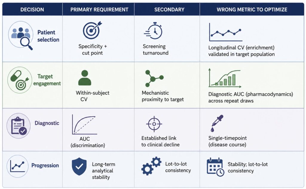
Patient selection asks a binary question at baseline: does this person have the pathology the drug targets? The assay needs high specificity and a defensible cut point, and it needs to return a result inside the screening window. Longitudinal precision is irrelevant.
Target engagement asks whether the drug reached its target and changed something measurable, usually within weeks. The assay needs low within-subject variability across repeat draws, and the analyte must sit close enough to the mechanism that a change is interpretable. Diagnostic accuracy is beside the point.
Progression tracking asks whether the disease course bent over months to years. The assay needs long-term analytical stability, a known relationship to clinical decline, and resistance to drift in reagent lots and site handling.
These requirements conflict. A marker with excellent baseline discrimination may be nearly static over 12 months and therefore useless as a pharmacodynamic readout. A marker that moves quickly may move for reasons unrelated to central nervous system pathology. Choosing one panel to serve all three purposes generally produces a panel that serves none of them well.
Figure 1. Biomarker selection by decision type
The biological definition changed what enrichment means
The field has moved from defining Parkinson’s disease clinically to defining it biologically. The Neuronal Synuclein Disease framework and its integrated staging system propose that misfolded neuronal α-synuclein plus dopaminergic dysfunction constitute the disease, with clinical syndrome treated as consequence rather than definition [2].
That reframing matters for enrollment because it exposes how heterogeneous “early Parkinson’s” has been. When the staging system was applied to baseline data from an observational cohort and two interventional trials, 1,030 of 1,741 participants with available seed amplification results were positive for pathological α-synuclein. Among sporadic Parkinson’s participants, 683 of 736 — about 93% — met the biological criteria. Genetic subgroups diverged sharply: roughly 64% of LRRK2 Parkinson’s participants and 33% of PRKN Parkinson’s participants were positive [3].
Enroll a LRRK2-enriched cohort without biological screening and a substantial minority of participants may not carry the pathology the drug engages. No dose adjustment recovers that.
The staging data also carry a power implication. Among participants in one sporadic cohort, median time to a clinically meaningful progression milestone was 8.3 years for baseline stage 2B, 5.9 years for stage 3, and 2.4 years for stage 4 [3]. Cohorts that mix these stages mix progression rates that differ by more than threefold. Biologically defined, functionally staged enrollment narrows that variance — which is the practical argument for biomarker-based selection, independent of any mechanistic claim.
Seed amplification assays have anchored this framework using cerebrospinal fluid, with reported sensitivity of 87.7% and specificity of 96.3% in a large multi-site cohort [1]. The performance is not in dispute. The access problem is: the framework’s own authors acknowledge feasibility and scalability limits of a CSF-dependent screening step and anticipate migration to more accessible matrices [3]. A trial that needs to screen several hundred candidates to enroll one hundred cannot readily do so through lumbar puncture.
What each blood marker class can and cannot tell you
Blood-based options in Parkinson’s fall into three broad classes, and conflating them is a common source of misspecified panels.
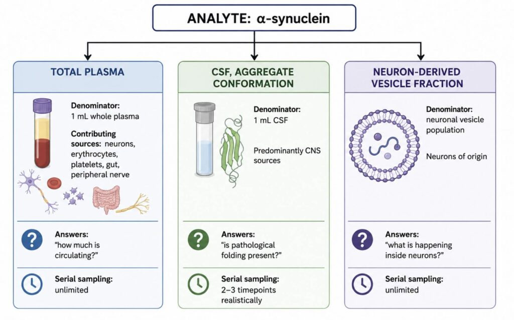
Total-plasma neurodegeneration markers. Serum neurofilament light chain rises in Parkinson’s relative to controls, increases with age and over time, and correlates with motor severity [6]. It is the most operationally mature blood biomarker available. It is also non-specific: the validating authors described its specificity for Parkinson’s as low, positioning it as useful for distinguishing Parkinson’s from atypical parkinsonism and for tracking progression rather than for defining disease [6]. Compounding this, plasma concentrations correlate negatively with body mass index and blood volume, so two patients with identical central pathology can return different values [7]. That is not an assay defect — it is a property of measuring a diluted analyte across a whole-body compartment.
Aggregate-conformation assays. Seed amplification detects pathological protein folding rather than protein abundance, which is why it discriminates so well. Applied to blood, the approach requires first concentrating a neuron-restricted fraction, then amplifying seeds from it.
Neuron-derived vesicle markers. Extracellular vesicles released by neurons cross into peripheral circulation carrying cargo that reflects the state of the originating cell. Isolating that population before measurement changes the denominator: instead of asking how much analyte is present per millilitre of plasma, the question becomes how much is present per neuron-derived vesicle population. Pathological α-synuclein has been detected in neuron-derived vesicles from blood and amplified by seeding assay [8]. Vesicle-associated α-synuclein in serum distinguished at-risk individuals with high prodromal probability from controls and correctly identified 80% of those who later phenoconverted, in a cross-sectional study of 576 participants [10]. Serum neuronal vesicle measurements have also differentiated Parkinson’s from atypical parkinsonian syndromes [11], and seeding activity in this compartment has been examined against disease duration [9]. A meta-analysis of central-nervous-system-originating vesicles across parkinsonian disorders found elevated α-synuclein in the neuronal fraction while flagging heterogeneity across isolation methods [15].
Figure 2. What the same analyte means in different compartments
Provenance, not sensitivity, is the axis that decides panel composition
The instinct when a blood signal is weak is to reach for a more sensitive detector. Digital immunoassay methods delivered exactly that over the past decade, pushing detection into the femtomolar range and making plasma neurodegeneration markers measurable at all. Before those methods existed, the argument about which compartment to measure was largely academic, because whole-plasma concentrations of most central nervous system analytes sat below the floor of available assays.
Sensitivity was the right problem to solve. It was not the only one. Measure α-synuclein in whole plasma with extraordinary precision and the result still aggregates neuronal, erythrocyte, platelet, enteric, and peripheral nerve contributions into a single number. The measurement is accurate. Its biological referent is ambiguous. For a target-engagement readout — where the question is specifically whether a drug changed something inside neurons — ambiguity about tissue of origin is disqualifying in a way that a modest loss of analytical sensitivity is not.
This is why panel composition should be settled on provenance grounds before sensitivity is discussed. Two practical consequences follow. First, a pharmacodynamic panel and a progression panel may legitimately draw on different compartments in the same trial, with a total-plasma neurodegeneration marker tracking long-term course while a neuron-restricted panel reads mechanism. Second, mechanism-adjacent readouts become available in the neuronal compartment that whole plasma cannot support. Presynaptic protein markers measured in plasma vesicles have been associated with motor progression in a cohort of 101 Parkinson’s participants and 43 controls, with baseline levels in the highest quartile predicting greater worsening on activities-of-daily-living and gait subscores [13]. Signal of that kind speaks to synaptic integrity rather than bulk axonal loss.
Build the panel against the trial objective
The following structure holds across most disease-modification programs.
Screening and enrichment. Establish biological eligibility. Where a program targets α-synuclein pathology directly, biological confirmation should be a screening criterion rather than a post-hoc subgroup variable. The distinction is not cosmetic: a subgroup analysis in a diluted cohort can only ever generate a hypothesis, while a screened cohort generates an answer. Blood-based conformational assays make repeat and large-scale screening tractable in a way CSF does not, which matters most in prodromal and at-risk populations where screen-failure rates are high and the consented population is asked to accept procedure burden before any prospect of benefit. Prespecify the cut point and the population it was validated in; a threshold established in clinically manifest disease may not transfer to a prodromal cohort, and a threshold that shifts after unblinding is not a threshold.
Target engagement. Select analytes with a defensible mechanistic link to the drug’s action, measured in the neuronal compartment, at intervals dense enough to distinguish drug effect from biological noise. Establish the within-subject coefficient of variation in a pilot before committing to a sampling schedule. This is the single most common omission — sponsors specify timepoints, then discover the assay’s repeat-measure variability exceeds the effect they hoped to detect. Two or three replicate baseline draws in a small cohort, spaced over the interval the trial will actually use, is usually enough to establish whether the plan is viable. Where it is not, the options are more frequent sampling, a larger cohort, or a different analyte, and it is considerably cheaper to learn that before randomization than after.
Progression and long-term course. Total-plasma neurodegeneration markers are appropriate here, with covariate adjustment for body mass index and renal function planned in the statistical analysis plan rather than added later [7]. Where the program’s hypothesis concerns synaptic preservation, add neuron-restricted synaptic readouts alongside.
Genetic subgroups. For GBA and LRRK2 cohorts, biological positivity rates differ enough from sporadic Parkinson’s that stratification variables should be defined at randomization [3]. This applies whether or not the drug is genotype-directed.
Validation and handling determine whether any of it holds
A biomarker plan is only as defensible as its pre-analytical control, and the neuronal vesicle literature has already been challenged on this point. A 2024 viewpoint in Movement Disorders argued that several studies applying seeding assays to blood-derived neuronal vesicles reported insufficient vesicle characterization — omitting tetraspanin marker panels and particle sizing after protocol modifications — and did not consistently reference ISEV reporting standards [12]. Whatever one concludes about the specific studies, the critique identifies the right failure mode. Two sensitive and modification-prone steps in series, vesicle enrichment followed by amplification, produce results that are difficult to reproduce across laboratories unless both steps are documented and controlled.
For a sponsor, that translates into concrete requests before samples are committed:
- Characterization data for the isolated vesicle population, reported against MISEV2023 recommendations, including tetraspanin marker evidence and particle size distribution [14]
- Within-run and between-run precision generated on the intended matrix, in the intended patient population, at concentrations spanning the expected range
- Documented stability across the actual collection-to-freezer interval at participating sites, including freeze-thaw tolerance
- Lot-to-lot bridging data, and a plan for what happens when a critical reagent lot changes mid-study
- Pre-specified handling requirements — tube type, processing window, centrifugation, aliquot volume — issued to sites as a manual of procedures, not as a protocol appendix
Sites will deviate. The question is whether deviations are detectable and quantifiable when they do.
Where this is heading
The biological definition of Parkinson’s disease is now established in research use, and the constraint on applying it at scale is matrix access rather than assay performance. Migration of conformational and mechanistic readouts from cerebrospinal fluid into blood is the near-term development that will determine how quickly biologically defined trials become routine rather than exceptional. Standardization is the gating factor: the field needs multi-site reproducibility data on neuron-restricted assays, generated under harmonized handling, before regulators will accept these measures as anything more than exploratory. Sponsors designing trials now can accelerate that by treating biomarker qualification as a deliverable of the program rather than a byproduct of it — prespecifying context of use, banking samples under controlled conditions, and publishing analytical performance alongside clinical results.
NeuroDex supports blood-based central nervous system biomarker programs using the ExoSORT™ platform to isolate neuron-derived extracellular vesicles from plasma, with panel design and analytical validation scoped to the decision the biomarker has to support.
References
[1] Siderowf A, Concha-Marambio L, Lafontant DE, et al. Assessment of heterogeneity among participants in the Parkinson’s Progression Markers Initiative cohort using α-synuclein seed amplification: a cross-sectional study. Lancet Neurol. 2023;22(5):407–417. https://doi.org/10.1016/S1474-4422(23)00109-6
[2] Simuni T, Chahine LM, Poston K, et al. A biological definition of neuronal α-synuclein disease: towards an integrated staging system for research. Lancet Neurol. 2024;23(2):178–190. https://doi.org/10.1016/S1474-4422(23)00405-2
[3] Dam T, Pagano G, Brumm MC, et al. Neuronal alpha-Synuclein Disease integrated staging system performance in PPMI, PASADENA, and SPARK baseline cohorts. npj Parkinsons Dis. 2024;10:178. https://doi.org/10.1038/s41531-024-00789-w
[4] Pagano G, Taylor KI, Anzures-Cabrera J, et al. Trial of prasinezumab in early-stage Parkinson’s disease. N Engl J Med. 2022;387(5):421–432. https://doi.org/10.1056/NEJMoa2202867
[5] Lang AE, et al. Trial of cinpanemab in early Parkinson’s disease. N Engl J Med. 2022;387(5):408–420. https://doi.org/10.1056/NEJMoa2203395
[6] Mollenhauer B, Dakna M, Kruse N, et al. Validation of serum neurofilament light chain as a biomarker of Parkinson’s disease progression. Mov Disord. 2020;35(11):1999–2008. https://doi.org/10.1002/mds.28206
[7] Manouchehrinia A, Piehl F, Hillert J, et al. Confounding effect of blood volume and body mass index on blood neurofilament light chain levels. Ann Clin Transl Neurol. 2020;7(1):139–143. https://doi.org/10.1002/acn3.50972
[8] Kluge A, Bunk J, Schaeffer E, et al. Detection of neuron-derived pathological alpha-synuclein in blood. Brain.2022;145(9):3058–3071. https://doi.org/10.1093/brain/awac115
[9] Schaeffer E, Kluge A, Schulte C, et al. Association of misfolded α-synuclein derived from neuronal exosomes in blood with Parkinson’s disease diagnosis and duration. J Parkinsons Dis. 2024;14(4):667–679. https://doi.org/10.3233/JPD-230390
[10] Yan S, Jiang C, Janzen A, et al. Neuronally derived extracellular vesicle α-synuclein as a serum biomarker for individuals at risk of developing Parkinson disease. JAMA Neurol. 2024;81(1):59–68. https://doi.org/10.1001/jamaneurol.2023.4398
[11] Jiang C, Hopfner F, Katsikoudi A, et al. Serum neuronal exosomes predict and differentiate Parkinson’s disease from atypical parkinsonism. J Neurol Neurosurg Psychiatry. 2020;91(7):720–729. https://doi.org/10.1136/jnnp-2019-322588
[12] Bernhardt AM, Nemati M, Boros FA, et al. α-Synuclein seed amplification assays from blood-based extracellular vesicles in Parkinson’s disease: an evaluation of the evidence. Mov Disord. 2024;39(8):1269–1271. https://doi.org/10.1002/mds.29923
[13] Hong CT, Chung CC, Yu RC, Chan L. Plasma extracellular vesicle synaptic proteins as biomarkers of clinical progression in patients with Parkinson’s disease. eLife. 2024;12:e87501. https://doi.org/10.7554/eLife.87501
[14] Welsh JA, Goberdhan DCI, O’Driscoll L, et al. Minimal information for studies of extracellular vesicles (MISEV2023): from basic to advanced approaches. J Extracell Vesicles. 2024;13(2):e12404. https://doi.org/10.1002/jev2.12404
[15] Taha HB, et al. Evaluation of α-synuclein in CNS-originating extracellular vesicles for Parkinsonian disorders: a systematic review and meta-analysis. CNS Neurosci Ther. 2023;29. https://doi.org/10.1111/cns.14341


Pingback: Can Blood-Based CNS Biomarkers Improve CNS Trial Endpoints?