



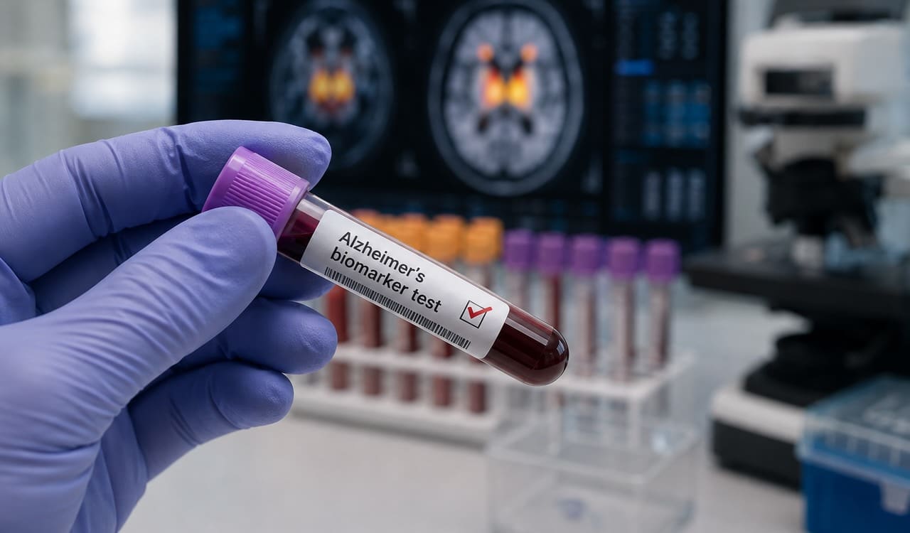
Biomarker Discovery for Alzheimer’s Drug Programs: What Blood-Based Platforms Can Add
Lecanemab and donanemab proved that amyloid-clearing drugs can slow decline — and proved something else along the way: confirming a patient’s amyloid status at baseline is not the same as tracking what a drug is doing to the brain across dosing. Between PET scans and lumbar punctures, sponsors are left inferring mechanism of action rather than measuring it. For programs now stacking anti-tau, anti-inflammatory, and combination strategies on top of amyloid clearance, that gap is becoming the rate-limiting step in translational biomarker strategy.
What Can Blood-Based Biomarkers Add to Alzheimer’s Drug Programs?
Blood-based CNS biomarkers give Alzheimer’s drug programs a non-invasive way to track mechanism of action between PET and CSF visits. Neuron-derived extracellular vesicles and neuroinflammation markers like GFAP add pathway-specific signal — synaptic, autophagy, and astrocyte-reactivity data — that neither imaging nor CSF was designed to capture on a dense sampling schedule.
Key Takeaways
- PET and CSF confirm amyloid or tau pathology at entry but were not built to track mechanism-of-action or pathway engagement between visits [1,2].
- Neuron-derived extracellular vesicles (NDEVs) carry synaptic, autophagy, and clearance-pathway cargo that can be sampled from plasma repeatedly and non-invasively [3,4,5].
- Astrocyte reactivity markers like GFAP add a neuroinflammation axis that complements — rather than duplicates — amyloid and tau readouts [9].
- Discovery-stage AD programs (combination MOA panels, preclinical-to-clinical bridging, at-risk cohort screening) are where blood-based EV platforms currently add the most value, ahead of any regulatory qualification pathway.
- Panel flexibility and documented reproducibility, not marketing claims, are what should differentiate a biomarker discovery partner for this stage of work.
The MOA Gap Anti-Amyloid and Anti-Tau Programs Still Face
Appropriate-use recommendations for lecanemab require a positive amyloid biomarker — PET or CSF p-tau181/Aβ42 — before treatment starts [1]. Donanemab’s TRAILBLAZER-ALZ program used the same logic, pairing amyloid PET with a tau PET stratification step to select and monitor patients through 76 weeks of dosing [2]. These are diagnostic and eligibility tools. They confirm that a patient has the pathology a drug is designed to treat, and they can show that a drug removed plaque. What they were never designed to answer is the question a translational team actually needs answered mid-trial: is the drug engaging its target and producing the downstream biological effect the mechanism predicts, in this patient, right now, between the imaging visits?
That distinction matters more as the field moves past single-mechanism amyloid clearance. Combination trials pairing anti-amyloid antibodies with anti-tau agents, anti-inflammatories, or metabolic modulators need to show that each arm’s specific mechanism is engaged — not just that amyloid went down. A sponsor running a combination arm with an anti-inflammatory compound, for instance, has no way to know from PET or CSF alone whether the inflammatory pathway actually moved, or whether any observed clinical benefit is attributable to the amyloid-clearing backbone alone. PET and CSF, sampled at most a handful of times per patient, cannot resolve that kind of arm-specific attribution. A biomarker capable of reflecting neuron-specific and pathway-specific biology on a denser sampling schedule can, and that gap between what regulators require for eligibility and what a translational team needs for internal go/no-go decisions is exactly where discovery-stage biomarker work now sits.
This is not an argument that CSF and PET should be replaced. Regulatory pathways for AD therapeutics are built around them, and that isn’t changing on the timeline of any current program. It’s an argument that treating them as the only tools available for tracking biology across a trial leaves real information on the table — information a plasma-based assay run between imaging visits could supply at a fraction of the burden.
Why CSF and PET Weren’t Built for Continuous MOA Monitoring
Two related, well-documented problems narrow what CSF and PET can tell a translational team.
First, sampling frequency. Lumbar puncture and PET are logistically and financially demanding enough that most protocols schedule two or three timepoints across a trial. A biological process that unfolds over weeks — receptor engagement, downstream signaling, a compensatory inflammatory response — will alias badly against that few observations.
Second, discordance. CSF and PET amyloid measures agree most of the time, but not always: cross-cohort work has found meaningful rates of discordant CSF-PET amyloid classification, with real consequences for who gets classified as a treatment candidate and how a trial’s eligibility criteria perform in practice [10]. When the two reference-standard measures don’t always agree with each other, using either alone as evidence of pathway-level mechanism of action — something neither was designed to assess — is asking more of the assay than its validation supports.
There’s a third, quieter problem: neither CSF nor PET has much to say about biology outside the amyloid/tau axis. Synaptic integrity, autophagy and lysosomal clearance, astrocyte reactivity, and metabolic signaling all play documented roles in AD progression, but none of them show up on an amyloid PET scan or in a standard CSF p-tau/Aβ42 panel. A translational team investigating a mechanism outside that axis — which describes most of the pipeline behind first-generation amyloid antibodies — has no diagnostic-grade blood or imaging tool built for the question they’re actually asking.
None of this is a defect in CSF or PET; both technologies do what they were built to do extremely well, and both remain the regulatory backbone for AD drug approval. The problem is that translational teams have started asking diagnostic-grade, infrequent-sampling tools calibrated to a single pathological axis to serve as pharmacodynamic-grade, dense-sampling, multi-pathway ones. That’s a mismatch between tool and task, not a flaw in either tool.
What Neuron-Derived Extracellular Vesicles Add to the Picture
Neuron-derived extracellular vesicles are membrane-bound particles shed by neurons that cross into peripheral blood and carry a molecular snapshot of their cell of origin — synaptic proteins, tau species, and components of the autophagy and lysosomal clearance machinery [3]. Because they can be isolated from a standard plasma draw, they support a sampling cadence — weekly, biweekly, whatever the protocol calls for — that CSF and PET cannot match without unacceptable patient burden.
The biomarker literature on NDEVs in Alzheimer’s has moved from proof-of-concept to defined analyte panels over the past three years. Synaptic proteins carried in plasma NDEVs have been proposed and clinically piloted as AD biomarkers [3], and a related study identified NMDAR2A as a marker of CNS-derived plasma EVs, building an integrative model that distinguished AD from healthy controls and Parkinson’s disease with strong discrimination in both discovery and validation cohorts [4]. NDEV cargo has also been shown to track cognitive decline longitudinally [6] and to shift measurably in response to an intervention as simple as structured exercise — evidence that these markers respond to biological change, not just static disease state [5].
A newer line of work extends NDEV cargo beyond synaptic and tau biology into brain insulin signaling, an AD-relevant pathway that has no other blood-accessible readout [7]. Brain insulin-signaling dysregulation is a documented feature of AD pathogenesis, but until this kind of NDEV work, there was no way to observe it in a living patient at all — it was a finding confined to post-mortem tissue. That illustrates the broader point for discovery-stage programs: NDEV panels are not a single-marker replacement for p-tau or amyloid. They’re a route to pathway-specific cargo — autophagy, synaptic integrity, insulin signaling, and more — that CSF and PET were never positioned to reach at all, independent of the sampling-frequency problem.
Reproducibility across isolation methods matters here as much as which analytes get measured. A comparison of isolation approaches for human brain-derived EVs found that yield and cargo composition can shift meaningfully depending on the method used to separate neuron-derived particles from the broader circulating EV pool [11]. For a discovery-stage program, that means the choice of isolation platform isn’t a back-office methods decision — it can determine whether a real biological signal survives sample processing intact, or gets diluted below the level of detection before it ever reaches the assay.
Neuroinflammation Signal Alongside Neuronal Cargo
Reactive astrocytosis, measured through plasma GFAP, has been shown to rise as early as ten years before the onset of cognitive impairment in individuals who go on to develop AD, tracking the severity of neuritic plaques and neurofibrillary tangles at autopsy [9]. In that same longitudinal cohort, GFAP elevation preceded the other blood biomarker changes examined, positioning astrocyte reactivity as an early rather than a downstream event in the biomarker cascade [9].
That’s a neuroinflammation axis running in parallel to amyloid and tau biology, not simply downstream of it. For combination programs pairing an anti-amyloid agent with an anti-inflammatory or microglial-modulating compound, having a neuron-derived readout and an astrocyte-reactivity readout from the same plasma draw gives a translational team two independent, mechanistically distinct axes instead of one — and because both come from the same standard plasma sample, adding the second axis doesn’t add a second patient visit or a second invasive procedure to the protocol.
Discovery-Stage Use Cases Where This Matters
Three situations come up repeatedly in AD programs where blood-based EV biomarker discovery earns its place well before any regulatory biomarker-qualification conversation starts:
Combination-therapy MOA panels. When a trial pairs mechanisms — amyloid clearance plus tau-directed therapy, or amyloid clearance plus a neuroinflammation-targeted compound — sponsors need to show each arm is doing its specific job, not just that the combined regimen produced a composite clinical or imaging effect. A synaptic/tau NDEV panel run alongside a GFAP readout gives two mechanistically distinct signals from one sample, at a cadence dense enough to build a real dose-response or time-course curve rather than three widely spaced data points.
Preclinical-to-clinical bridging. Biomarker signals discovered in animal models don’t always hold up in human plasma — a persistent failure mode in translational neuroscience generally, not just in AD. NDEV and astrocyte-marker panels that have already been characterized in both transgenic mouse models and human cohorts give translational teams a documented starting point for checking whether a preclinical signal is likely to translate, rather than discovering the mismatch after an expensive Phase I/II endpoint decision has already been locked in [9]. That bridging step is where a discovery-stage biomarker partner can do the most good relative to cost: it’s far cheaper to find out a marker doesn’t translate during IND-enabling work than during a dosed human trial.
At-risk and genetic-cohort screening. Prevention trials in cognitively unimpaired, genetically high-risk populations — APOE ε4 homozygotes, autosomal-dominant AD kindreds, and similar cohorts — need a low-burden way to screen and monitor large numbers of participants over years, often before any symptoms appear and well before a positive PET or CSF result would be expected. Blood draws scale in a way that serial PET and lumbar puncture do not, both financially and in terms of what a healthy volunteer is willing to tolerate repeatedly over a multi-year prevention protocol. NDEV-based discovery work in this space is still building toward full population-scale deployment, but the sampling logic already fits the use case better than imaging or CSF, and early characterization work in this area is where sponsors are currently investing.
What to Demand From a Discovery-Stage Biomarker Partner
Not every EV assay is built the same way, and the differences matter more at the discovery stage than they might later, because a weak signal here can sink a program before it reaches a pivotal trial.
Isolation and characterization methods should be reported against the field’s current consensus standard — MISEV2023 — rather than against an internal, unpublished protocol; the guideline exists specifically because EV isolation methods vary enough to change what gets measured [8]. A comprehensive comparison of isolation approaches for human brain-derived EVs found meaningful differences in yield and cargo composition depending on method, which is exactly the kind of variability a discovery-stage partner should be able to speak to directly, not gloss over [11].
Beyond methodology, four practical questions are worth asking before committing samples to a platform.
Can the panel be customized to the specific pathway your mechanism engages, rather than forcing your program onto a fixed marker list built for a different indication? A combination trial targeting neuroinflammation needs different cargo readouts than one targeting synaptic loss, and a partner offering only one fixed panel is optimizing for their own operational simplicity, not your program’s biology.
What is the documented coefficient of variation across replicate plasma samples, and over what sample volume? Reproducibility numbers that only exist in an internal validation deck, without a published or at least externally reviewed comparison, are hard to weigh against a competing platform’s claims.
Can the partner isolate neuron-derived vesicles specifically, distinguishing neuronal cargo from the broader pool of circulating EVs, without requiring more plasma than your protocol has budgeted per timepoint? Sample volume constraints are often set early in protocol design, sometimes before a biomarker vendor is even selected, and a platform that needs more plasma than the protocol allows is a non-starter regardless of how good its assay is on paper.
And finally: does the partner report their isolation and characterization methodology against a recognized field standard, or only against their own internal benchmarks? Given how much isolation method affects yield and cargo composition [11], a partner unwilling or unable to describe their method against MISEV2023 terminology is asking you to trust a black box.
NeuroDex’s ExoSORT™ platform isolates neuron-derived EVs from standard plasma volumes for exactly this kind of discovery-stage work, with panels built around the pathway a program needs to track rather than a fixed marker menu. More detail on validation approach is at neurodex.co.
Where This Is Headed
The near-term trajectory for blood-based AD biomarkers runs through better-characterized multi-analyte panels — NDEV cargo alongside GFAP, p-tau, and NfL — rather than any single marker displacing CSF or PET outright. Large-scale proteomic and multi-omic consortia are already assembling cross-cohort datasets at a scale individual sponsors couldn’t build alone, which will accelerate which NDEV and neuroinflammation markers hold up across populations. For discovery-stage translational teams, the near-term opportunity isn’t waiting for that consolidation to finish — it’s using a flexible, well-characterized EV platform now to get mechanism-of-action data that PET and CSF were never going to provide, at a sampling cadence a trial can actually afford.
References
[1] Cummings J, Apostolova L, Rabinovici GD, et al. Lecanemab: Appropriate use recommendations. J Prev Alzheimers Dis. 2023;10(3):362-377. doi:10.14283/jpad.2023.30. https://www.jpreventionalzheimer.com/7041-lecanemab-appropriate-use-recommendations.html
[2] Shcherbinin S, Evans CD, Lu M, et al. Association of amyloid reduction after donanemab treatment with tau pathology and clinical outcomes: the TRAILBLAZER-ALZ randomized clinical trial. JAMA Neurol. 2022;79(10):1015-1024. doi:10.1001/jamaneurol.2022.2793. https://pmc.ncbi.nlm.nih.gov/articles/PMC9468959/
[3] Eitan E, Thornton-Wells T, Elgart K, et al. Synaptic proteins in neuron-derived extracellular vesicles as biomarkers for Alzheimer’s disease: novel methodology and clinical proof of concept. Extracell Vesicles Circ Nucl Acids. 2023;4(1):133-150. doi:10.20517/evcna.2023.13. https://www.oaepublish.com/articles/evcna.2023.13
[4] Tian C, et al. Blood extracellular vesicles carrying synaptic function- and brain-related proteins as potential biomarkers for Alzheimer’s disease. Alzheimers Dement. 2023. doi:10.1002/alz.12723. https://alz-journals.onlinelibrary.wiley.com/doi/10.1002/alz.12723
[5] Delgado-Peraza F, Nogueras-Ortiz C, Simonsen AH, et al. Neuron-derived extracellular vesicles in blood reveal effects of exercise in Alzheimer’s disease. Alzheimers Res Ther. 2023;15:156. doi:10.1186/s13195-023-01303-9. https://pmc.ncbi.nlm.nih.gov/articles/PMC10510190/
[6] Eren E, Leoutsakos JM, Troncoso J, Lyketsos CG, Oh ES, Kapogiannis D. Neuronal-derived EV biomarkers track cognitive decline in Alzheimer’s disease. Cells. 2022;11(3):436. doi:10.3390/cells11030436. https://pmc.ncbi.nlm.nih.gov/articles/PMC8834433/
[7] Cleary JP, et al. Neuron-derived extracellular vesicles as a liquid biopsy for brain insulin dysregulation in Alzheimer’s disease and related disorders. Alzheimers Dement. 2025. doi:10.1002/alz.14497. https://alz-journals.onlinelibrary.wiley.com/doi/full/10.1002/alz.14497
[8] Welsh JA, Goberdhan DCI, O’Driscoll L, et al. Minimal information for studies of extracellular vesicles (MISEV2023): from basic to advanced approaches. J Extracell Vesicles. 2024;13(5):e12404. doi:10.1002/jev2.12404. https://pmc.ncbi.nlm.nih.gov/articles/PMC10850029/
[9] Varma VR, An Y, Kac PR, et al. Longitudinal progression of blood biomarkers reveals a key role of reactive astrocytosis in preclinical Alzheimer’s disease. Med. 2025;Jun 3:100724. doi:10.1016/j.medj.2025.100724. https://www.sciencedirect.com/science/article/abs/pii/S2666634025001515
[10] de Wilde A, Reimand J, Teunissen CE, et al. Discordant amyloid-β PET and CSF biomarkers and its clinical consequences. Alzheimers Res Ther. 2019;11:78. doi:10.1186/s13195-019-0532-x. https://pmc.ncbi.nlm.nih.gov/articles/PMC6739952/
[11] Zhang Z, Yu K, You Y, et al. Comprehensive characterization of human brain-derived extracellular vesicles using multiple isolation methods: implications for diagnostic and therapeutic applications. J Extracell Vesicles. 2023;12(8):e12358. doi:10.1002/jev2.12358. https://pmc.ncbi.nlm.nih.gov/articles/PMC10415636/


Leave a Reply