



TDP-43 in Blood: Proving Target Engagement
A biotech team advancing a TDP-43-targeting ASO into first-in-human dosing faces a familiar problem: the drug lowers pathogenic TDP-43 in animal models, but the sponsor has no scalable way to confirm that same effect in patients without asking them to return for repeated lumbar punctures. Dose-finding depends on it. So does the go/no-go decision before a Phase II commitment. The biology of the target isn’t the bottleneck — the biomarker is.
Key Takeaways
- TDP-43-targeting therapeutics (small-molecule stabilizers, ASOs, and targeted degraders) need repeatable, blood-based pharmacodynamic proof of target engagement to support dose-finding and go/no-go decisions.
- Total plasma TDP-43 has produced contradictory results across cohorts — elevated in some disease-enriched samples, unassociated with outcomes in general population cohorts — because peripheral, non-neuronal TDP-43 dilutes the signal [1,2,3].
- Neuron-derived extracellular vesicle (NDEV) isolation restricts the signal to CNS-origin cargo before quantification, addressing the specificity problem that limits whole-plasma TDP-43 assays [3,4].
- Precedent for blood- and CSF-based pharmacodynamic proof of target engagement already exists for other CNS-targeting ASOs, including a MAPT-targeting oligonucleotide that reduced CSF tau in a Phase 1b trial [5].
- A GLP-compliant NDEV isolation and quantification workflow can be built modality-agnostically, supporting ASOs, degraders, and small molecules with a shared blood-based readout across a dosing study.
Why TDP-43-Targeting Drugs Need a Different Kind of Biomarker
Modality diversity is one of the more encouraging developments in the TDP-43 therapeutic space. Antisense oligonucleotides directed at upstream regulators, targeted protein degraders aimed at clearing pathogenic aggregates, and small molecules intended to stabilize normal nuclear TDP-43 localization are all in various stages of preclinical and early clinical development for ALS, FTD, and LATE. Each of these modalities makes a distinct, testable prediction about what should change in the patient’s biology after dosing — reduced pathogenic aggregate burden, restored nuclear localization, or altered downstream RNA splicing products.
The problem is translating that prediction into a measurement a sponsor can actually collect. CSF sampling supports two, maybe three, timepoints per patient across a dose-escalation study before patient burden and site logistics become limiting. That’s workable for a single confirmatory sample. It’s not workable for the kind of dense pharmacodynamic curve a modality-specific program needs to distinguish a dose-dependent effect from noise, particularly in early-phase studies where the sponsor is still learning the dose-response relationship.
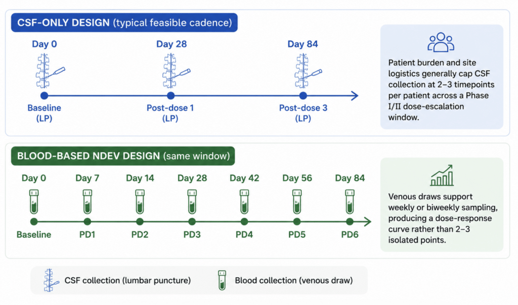
Figure 1. Sampling density: CSF-only vs. blood-based pharmacodynamic monitoring across a dose-escalation study
What CSF and NfL Already Tell You — and Where They Stop
CSF-based TDP-43 measurement has legitimate value, and a systematic review and meta-analysis found CSF TDP-43 levels are elevated in ALS patients relative to controls across seven pooled studies, supporting its candidacy as a disease-state marker [1]. Neurofilament light chain (NfL), meanwhile, remains the most clinically validated blood-based marker of neurodegeneration in ALS and is widely used to track neuroaxonal injury over the course of a trial.
Both of these are useful. Neither answers the specific question a TDP-43-targeting program needs answered: did the drug engage TDP-43 biology, and by how much, at this dose, in this patient? NfL reflects downstream neuronal injury broadly — it doesn’t distinguish a TDP-43-specific mechanism of action from any other source of axonal damage. CSF TDP-43 answers the mechanism question but not the sampling-frequency question; the same meta-analysis that supports CSF TDP-43’s diagnostic signal also notes that further studies are needed before firm conclusions can be drawn about its performance as a repeated-measures biomarker [1].
The Specificity Problem: Why Plasma TDP-43 Alone Hasn’t Delivered
The natural next step — measuring total TDP-43 directly in plasma rather than CSF — has produced a genuinely mixed evidence base, and it’s worth being direct about why rather than glossing over it.
In a population-based cohort of 1,058 participants from the Cardiovascular Health Study, plasma TDP-43 levels were not associated with cognitive decline, incident dementia, brain MRI volumes, or plasma AD biomarkers over a mean 5.5-year follow-up [2]. In a separate case-control comparison drawing on autopsy-confirmed LATE neuropathologic change, plasma TDP-43 and phospho-TDP-43 were significantly elevated in advanced disease, with an ROC area under the curve approaching 0.8 in the subgroup with comorbid Alzheimer’s pathology [3]. Both studies used well-characterized cohorts. The disagreement isn’t a data quality problem — it’s a specificity problem.
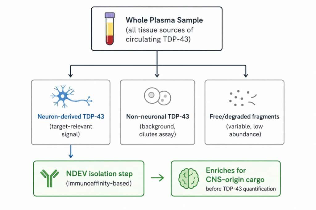
TDP-43 is expressed outside the CNS. A plasma sample captures TDP-43 shed from any tissue, and the neuron-derived signal a TDP-43-targeting drug is meant to affect gets diluted by everything else in circulation. That dilution behaves differently across cohorts depending on comorbidity burden, disease stage, and assay platform, which is a reasonable explanation for why population-based and disease-enriched cohorts have produced different conclusions using the same analyte [2,3].
Figure 2. Where the TDP-43 signal gets lost — and where NDEV isolation targets it
Neuron-Derived EVs as a Pharmacodynamic Window Into TDP-43 Biology
Brain-derived extracellular vesicles carry cargo — protein and RNA — that reflects the biology of the cell of origin at the time of release, and a 2024 review examining their use within the Research Domain Criteria (RDoC) framework specifically highlighted their potential as non-invasive mechanistic biomarkers of target engagement, treatment response, and toxicity in CNS drug development [4]. That review also flagged the standardization work still required across the field — isolation method, characterization approach, and cargo readout all need to be locked down before an NDEV assay can support regulatory-grade decisions [4].
This is where isolating the neuron-derived fraction before quantification changes the picture. Restricting the analyte pool to vesicles carrying neuronal surface markers removes much of the non-neuronal background that confounds whole-plasma TDP-43 measurement, addressing the same dilution problem responsible for the inconsistent cohort findings described above [2,3,4]. Improved immunoassay platforms for TDP-43 and its disease-associated modifications are also an active area of development in parallel, reflecting broader field recognition that detection sensitivity, not just isolation specificity, needs continued refinement [7].
None of this displaces MISEV2023 guidance on rigor in EV isolation and characterization — quite the opposite. A pharmacodynamic biomarker intended to support dosing decisions in a therapeutic program needs the same standardization discipline the guidelines call for: documented isolation method, defined vesicle markers, and validated recovery and reproducibility metrics before the assay is used to make a go/no-go call [8].
Designing a Target Engagement Strategy Across Modalities
The three TDP-43-targeting modalities in active development each need a slightly different pharmacodynamic readout, but all three can draw on the same NDEV isolation platform as the sample-prep layer.
| Modality | What “target engagement” looks like | What the NDEV readout needs to capture |
|---|---|---|
| Antisense oligonucleotides (ASOs) | Reduced pathogenic transcript/protein expression | Dose-dependent reduction in NDEV TDP-43 or its disease-associated phospho-forms over serial timepoints |
| Targeted protein degraders | Clearance of aggregated/misfolded TDP-43 | Reduction in aggregate-associated TDP-43 signal relative to baseline, tracked across dose cohorts |
| Small-molecule stabilizers | Restored nuclear localization, normalized splicing function | Change in TDP-43 cargo levels and, where feasible, downstream RNA splicing biomarkers within the same NDEV fraction |
Table is illustrative, built from general pharmacodynamic biomarker design principles for CNS drug development and modality-specific mechanisms of action [4,6].
There’s useful precedent here from a different CNS target. A tau-targeting antisense oligonucleotide (MAPT_Rx) produced dose-dependent reductions in CSF total tau and phospho-tau in a Phase 1b, randomized, placebo-controlled trial in mild Alzheimer’s disease, and those biomarker changes served as the pharmacodynamic evidence supporting the mechanism before the program advanced [5]. The trial design question for TDP-43-targeting programs is whether that same kind of dose-response evidence can be generated with fewer LP visits and a denser sampling schedule — which is the specific gap blood-based NDEV monitoring is positioned to close.
What GLP-Validated NDEV Biomarker Support Looks Like in Practice
For a sponsor moving a TDP-43-targeting ASO, degrader, or small molecule toward IND-enabling studies and into first-in-human dosing, the practical need is a biomarker partner that can take a candidate target, confirm assay compatibility with the specific analyte (whether that’s total TDP-43, a phospho-specific form, or a splicing-product surrogate), and validate detection sensitivity against the expected magnitude of treatment effect — all before the first patient is dosed.
NeuroDex’s ExoSORT™ platform isolates neuron-derived extracellular vesicles from plasma using an immunoaffinity-based enrichment step, producing the CNS-origin fraction that downstream TDP-43 immunoassays need in order to separate target-relevant signal from peripheral background. That isolation step is what makes it possible to run the same assay across a dense dose-escalation sampling schedule using blood draws instead of lumbar punctures, and to do so under GLP-compliant conditions suitable for supporting regulatory submissions.
The field’s TDP-43 evidence base is still being built out — the population-cohort findings described above are a legitimate reminder that whole-plasma measurement alone hasn’t settled the question [2,3]. What’s changing is the tooling available to separate signal from background before that question gets asked again.
References
[1] Gambino CM, Ciaccio AM, Lo Sasso B, Giglio RV, Vidali M, Agnello L, Ciaccio M. The role of TAR DNA binding protein 43 (TDP-43) as a candidate biomarker of amyotrophic lateral sclerosis: a systematic review and meta-analysis. Diagnostics (Basel). 2023;13(3):416. https://doi.org/10.3390/diagnostics13030416
[2] Fohner AE, Sitlani CM, Jayadev S, Bis JC, Trittschuh EH, Lopez OL, Tracy RP, Psaty BM, Longstreth WT Jr, Seshadri S. Plasma TAR DNA-binding protein 43 (TDP-43) levels in a population-based cohort of older adults: The Cardiovascular Health Study. J Alzheimers Dis. 2025;105(4):1275-1281. https://doi.org/10.1177/13872877251334820
[3] Wang J, Schneider JA, Bennett DA, Seyfried NT, Young-Pearse TL, Yang HS. Plasma TDP-43 is a potential biomarker for advanced limbic-predominant age-related TDP-43 encephalopathy neuropathologic change. Mol Neurodegener. 2025;20:119. https://doi.org/10.1186/s13024-025-00910-4
[4] Magaraggia I, Krauskopf J, Ramaekers JG, You Y, de Nijs L, Briedé JJ, Schreiber R. Harnessing brain-derived extracellular vesicles to support RDoC-based drug development. Neuroscience Applied. 2024;4:105406. https://doi.org/10.1016/j.nsa.2024.105406
[5] Mummery CJ, Börjesson-Hanson A, Blackburn DJ, Vijverberg EGB, De Deyn PP, Ducharme S, Jonsson M, Schneider A, Rinne JO, Ludolph AC, Bodenschatz R, Kordasiewicz H, Swayze EE, Fitzsimmons B, Mignon L, Moore KM, Yun C, Baumann T, Li D, Norris DA, Crean R, Graham DL, Huang E, Ratti E, Bennett CF, Junge C, Lane RM. Tau-targeting antisense oligonucleotide MAPT_Rx in mild Alzheimer’s disease: a phase 1b, randomized, placebo-controlled trial. Nat Med. 2023;29:1437-1447. https://doi.org/10.1038/s41591-023-02326-3
[6] Baljinnyam B, Coussens NP, Simeonov A. Editorial: Biophysical target engagement assays in chemical biology and pharmacological research. Front Cell Dev Biol. 2023;11:1163966. https://doi.org/10.3389/fcell.2023.1163966
[7] Demos C, Padmanabhan N, Uttarala S, Dzantiev L, Berry JD, Mathew A, Stengelin M, Sigal G, Wohlstadter JN. Novel immunoassays for TDP-43 detection in plasma and CSF. Alzheimers Dement. 2025 [ahead of print, conference abstract]. https://doi.org/10.1002/alz.70856
[8] Welsh JA, Goberdhan DCI, O’Driscoll L, et al. Minimal information for studies of extracellular vesicles (MISEV2023): From basic to advanced approaches. J Extracell Vesicles. 2024;13(2):e12404. https://doi.org/10.1002/jev2.12404
Leave a Reply