The α-Synuclein Blueprint: Understanding its Critical Role in Parkinson’s Disease Pathology
Parkinson’s disease (PD) is a complex and relentless neurodegenerative disorder that gradually erodes motor control and cognitive function, affecting millions of people across the globe. For decades, the precise molecular events triggering this decline remained a scientific enigma. However, a quarter-century of intensive research has increasingly converged on a single protein: alpha-synuclein (α-Synuclein). Normally a benign and functional component within the intricate machinery of our brain cells, this protein can undergo a devastating transformation, becoming a primary agent of cellular destruction. Understanding its journey from a helpful servant to a pathological master is central to deciphering the enigma of Parkinson’s disease. This article serves as a blueprint, mapping the intricate role of the synuclein protein from its native function to the cascade of events that culminates in neurodegeneration, and exploring the therapeutic strategies being developed to correct the flaws in its design.
Parkinson’s Disease: A Progressive Neurodegenerative Condition
As one of the most common neurodegenerative conditions, Parkinson’s disease is clinically defined by the progressive and selective loss of specific brain cells. The most profound damage occurs to the dopaminergic neurons—cells that produce the neurotransmitter dopamine—located in a critical midbrain region called the substantia nigra pars compacta. Dopamine is a key neurotransmitter for movement. Losing these dopamine neurons causes the main motor symptoms of PD, which include tremors, stiffness, slow movement (bradykinesia), and balance problems. As the disease advances, the pathology spreads beyond the motor circuits, manifesting in a wide range of non-motor symptoms, including cognitive impairment, sleep disorders, and a diminished sense of smell, sometimes progressing for years before the classic motor signs appear.
Introducing α-Synuclein: The Central Player in PD Pathology
At the molecular heart of Parkinson’s pathology lies the synuclein protein, α-Synuclein. This protein is abundantly expressed in the human brain, concentrated at the presynaptic termini—the communication hubs where one neuron sends signals to another. While its precise functions are still being fully elucidated, it is understood to play a crucial role in regulating the release of neurotransmitters. In Parkinson’s disease, this protein misfolds and forms toxic clumps. These clumps are the main part of Lewy bodies and Lewy neurites. These aggregates, found within surviving neurons, are the defining pathological signatures of PD and related disorders known as synucleinopathies or Lewy body diseases.

The “Blueprint” Metaphor: From Normal Function to Pathological Cascade
To truly grasp the disease process, we can conceptualize α-Synuclein’s genetic code—the SNCA gene—and its normally folded structure as its “original blueprint.” This blueprint dictates its proper function within the neuron, ensuring smooth synaptic communication and trafficking. In Parkinson’s, this blueprint becomes corrupted. Genetic mutations, environmental triggers, or chronic cellular stress can cause the protein to deviate from its intended structure. This initiates a catastrophic pathological cascade where the altered blueprint leads to misfolding, protein aggregation, and a chain reaction of cellular damage that ultimately drives the relentless neurodegeneration seen in PD.
Understanding the Synuclein Protein: Structure and Location
Alpha-synuclein is a small protein made of 140 amino acids. It is encoded by the SNCA gene. In its healthy, soluble native state within the cytosol of brain cells, it is considered “natively unfolded,” lacking a single, rigid three-dimensional structure. This inherent flexibility allows it to interact with a variety of partners, most notably the lipid membranes of synaptic vesicles. Its primary residence at the presynaptic termini places it in the perfect position to modulate neurotransmitter trafficking and release, acting as a key regulator in the delicate dance of neuronal communication.
Essential Roles in Neuronal Function
The original blueprint for α-Synuclein specifies several vital tasks essential for neuronal health. Its principal role involves the regulation of synaptic vesicles—the tiny lipid sacs that store and release neurotransmitters. By binding to these vesicles, α-Synuclein helps ensure they are readily available for release and modulates the efficiency of synaptic transmission. It contributes to the maintenance of the synaptic vesicle pool and interacts with key components of the cellular machinery, such as the SNARE protein ykt6, to facilitate protein trafficking and preserve the integrity of the synapse, ensuring clear and efficient communication between neurons.
From Native State to Misfolded Alpha-Synuclein
The journey from a functional protein to a pathogenic agent—a critical deviation from the blueprint—begins when α-Synuclein abandons its soluble, unfolded state. This process, central to the molecular pathogenesis of PD, involves the protein adopting a conformation rich in β-sheets. This structural transformation makes the protein “sticky,” causing it to self-associate and begin the process of aggregation. A multitude of factors can trigger this initial misfolding event, including mutations in the SNCA gene, which can lead to autosomal dominant PD, overproduction of the protein, or cellular insults that increase oxidative stress.
The Formation of Toxic Alpha-Synuclein Species
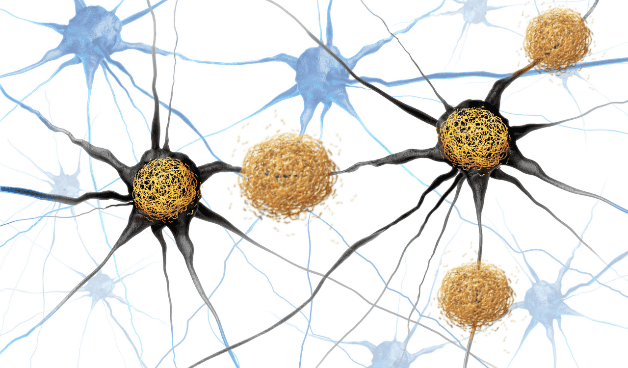
Once misfolded, individual α-Synuclein monomers begin to clump together in a process known as α-Syn aggregation. They first form small, soluble clusters called α-syn oligomers, which are now widely considered the most neurotoxic species, capable of permeating membranes and disrupting numerous cellular functions. These oligomers can further assemble into larger, thread-like protofibrils and, ultimately, into the large, insoluble amyloid fibrils that constitute the core of Lewy bodies and Lewy neurites found in post-mortem human brain tissue. This progressive accumulation often follows a predictable pattern described by the Braak stages, beginning in areas like the olfactory bulb and gut before ascending into the brain.
The Role of Post-Translational Modifications (PTMs)
The α-Synuclein blueprint can be further altered after the protein is synthesized through a variety of chemical changes known as post-translationally modified forms of αSyn. These modifications, such as phosphorylation, ubiquitination, nitration, and truncation, can dramatically influence the protein’s behavior. For instance, phosphorylation at the serine 129 residue—a modification detected by the widely used pSer129 antibody—is found on over 90% of aggregated α-Synuclein within Lewy bodies, compared to only about 4% of the total protein in a healthy brain, according to research in Molecular Neurodegeneration. Similarly, phosphorylation at tyrosine 39 (pTyr39) and C-terminal truncations, such as the formation of the αSyn species truncated at aa 119, can create a peptide fragment that enhances the protein’s propensity for aggregation and toxicity.
Cellular Mayhem: α-Synuclein’s Impact on Neuronal Function and Survival
The accumulation of misfolded α-syn oligomers and larger fibrils unleashes a storm of dysfunction within the neuron. This pathological protein acts not as a single-point failure but as a saboteur that compromises multiple interconnected systems. This disruption leads to a state of cellular mayhem that compromises neuronal health, impairs communication, and ultimately culminates in the death of vulnerable brain cells, particularly the dopamine neurons that are central to motor control.
Lysosomal Dysfunction: The Cell’s Compromised Waste Disposal System
Neurons depend on a sophisticated waste clearance system known as the autophagy-lysosomal pathway to degrade and recycle damaged proteins and organelles. Aggregated α-Synuclein directly sabotages this critical process, leading to severe lysosomal dysfunction. It can impair autophagosome formation, damage lysosomal membranes, and inhibit the activity of key degradative enzymes like Cathepsin D. This effectively clogs the cell’s recycling machinery, preventing the removal of toxic α-Synuclein and other cellular debris. This pathway is particularly relevant in the context of genetic risk factors for PD; for instance, GBA1 mutations, which cause the lysosomal storage disorder Gaucher disease, are the most common genetic risk factor for Parkinson’s, as they impair Lysosomal activity and create an environment ripe for α-Synuclein accumulation.
Mitochondrial Dysfunction: An Energy Crisis in Neurons
Mitochondria, the powerhouses of the cell, are exquisitely vulnerable to toxic α-Synuclein. As aggregates accumulate, they can interact directly with mitochondrial membranes and key proteins like the Mitochondrial pyruvate carrier, disrupting their integrity and impairing the electron transport chain. This leads to profound mitochondrial dysfunction and an energy crisis, especially in the highly active dopaminergic neurons of the substantia nigra pars compacta. Damaged mitochondria produce a lot of oxidative stress. They release harmful reactive oxygen species (ROS). These damage lipids, proteins, and DNA. This creates a harmful cycle of mitochondrial problems and cell damage.
Neuroinflammation: The Immune System’s Double-Edged Sword
The brain’s immune cells, microglia and astrocytes, are activated by signals of neuronal stress. When dying neurons release aggregated α-Synuclein, it acts as a danger signal, triggering chronic neuroinflammation through pathways like the NLRP3 inflammasome. This activation starts as a protection. It causes the release of pro-inflammatory cytokines and other toxic molecules. These can harm nearby neurons. This damage makes neurodegeneration worse in a cycle that keeps going. This sustained inflammatory state contributes significantly to the progressive nature of Parkinson’s disease.
Synaptic Impairment: Communication Breakdown in Brain Cells
Long before a neuron succumbs to cell death, its primary function—communication—is severely compromised. Misfolded α-Synuclein accumulates at presynaptic termini, where it disrupts its normal role in neurotransmitter release. This leads to impaired dopamine trafficking, faulty recycling of synaptic vesicles, and a breakdown in the structural integrity of the synapse. This synaptic dysfunction is one of the earliest events in the disease process, contributing significantly to both motor and non-motor symptoms well before widespread neuronal loss is evident in the substantia nigra pars compacta.
Prion-like Propagation: How Misfolded α-Synuclein Spreads
One of the most insidious features of misfolded alpha-synuclein is its ability to spread from one neuron to another in a prion-like manner. Toxic α-syn oligomers can be released from a compromised neuron and taken up by a neighboring healthy one, possibly through mechanisms like tunneling nanotubes. Once inside, these pathological “seeds” corrupt the healthy, native α-Synuclein in the recipient cell, templating its misfolding and kickstarting the aggregation process anew. This trans-neuronal propagation allows the pathology to spread through anatomically connected brain circuits, explaining the progressive and predictable pattern of symptom development in Parkinson’s disease.
Selective Neuronal Vulnerability and Neurodegeneration
While α-Synuclein is present throughout the neural tissue of the brain, the dopaminergic substantia nigra neurons are uniquely susceptible to its toxic effects. Several ideas explain why this happens. These include the neurons’ high energy needs, their long axonal networks, and the oxidative stress made during dopamine metabolism. This combination of factors creates a “perfect storm” of vulnerability, leading to their progressive death—neurodegeneration—which directly causes the debilitating motor symptoms of Parkinson’s.
The Gut-Brain Axis: A Gateway for α-Synuclein Propagation
A growing body of compelling evidence suggests that the pathological cascade of Parkinson’s may not begin in the brain at all. The gut-brain axis has emerged as a critical area of research, with the Braak hypothesis proposing that misfolded alpha-synuclein first appears in the neurons of the enteric nervous system (the gut) or the olfactory bulb. From these peripheral sites, the pathology could travel up nerve pathways, such as the vagus nerve, to enter the brainstem and subsequently spread throughout the brain. This theory provides a plausible explanation for some of the earliest non-motor symptoms of PD, like constipation and loss of smell, which can precede motor deficits by years.
Genetic Predisposition to Alpha-Synuclein Pathology
While most cases of Parkinson’s disease are considered sporadic, a subset is directly linked to genetic mutations that corrupt the α-Synuclein blueprint. Point mutations or multiplications of the SNCA gene can lead to the production of an α-Synuclein protein that is inherently more prone to aggregation or is simply overproduced, overwhelming the cell’s clearance capacity. In addition to SNCA, mutations in genes like LRRK2 and the GBA gene also significantly increase PD risk, often by intersecting with α-Synuclein-related pathways, such as lysosomal function and cellular stress responses. This genetic evidence provides the strongest causal link between α-Synuclein and the pathogenesis of PD.
Environmental Factors: Triggers and Modifiers of Disease Progression
It is widely believed that the development of sporadic PD involves a complex interplay between genetic predisposition and environmental factors. Chronic exposure to certain pesticides (e.g., paraquat, rotenone), industrial solvents, and heavy metals has been associated with an increased risk of developing the disease. These environmental toxins are thought to act as triggers, initiating the pathological cascade by inducing mitochondrial dysfunction, increasing oxidative stress, or directly promoting the protein aggregation of α-Synuclein, thereby corrupting the blueprint in genetically susceptible individuals.
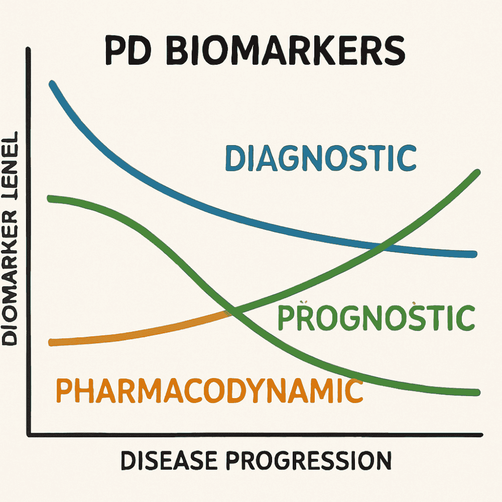
Biomarkers for Early Detection and Disease Progression Monitoring
A significant hurdle in treating Parkinson’s is the absence of definitive biomarkers for early and accurate diagnosis. By the time characteristic motor symptoms emerge, substantial and irreversible damage to the dopaminergic neurons has already occurred. Researchers are therefore intensely focused on developing biomarkers. Recently, seed amplification assays (SAAs) have shown great promise. They can detect tiny amounts of misfolded α-Synuclein in CSF with high accuracy. This could change early diagnosis and facilitate the clinical testing of new disease-modifying therapies.
Advanced Imaging and Tissue Analysis Techniques
Our understanding of the α-Synuclein blueprint has been dramatically advanced by sophisticated research tools. Analysis of formalin-fixed, paraffin-embedded brain tissue from brain banking programs allows scientists to visualize pathology directly. Techniques like confocal microscopy and super-resolution methods like stimulated emission depletion microscopy provide stunningly detailed images of Lewy bodies and their subcellular interactions. Multiplex immunofluorescence, using tools like phospho-Ser129 alpha-synuclein monoclonal antibodies, allows for single-cell quantification of multiple pathological markers. Furthermore, cutting-edge techniques like cry-electron microscopy, as detailed in journals like Molecular Neurodegeneration, are revealing the high-resolution atomic structure of α-Synuclein fibrils.
Disease-Modifying Therapies: Halting or Reversing Pathology
Current treatments for Parkinson’s primarily offer symptomatic relief; they do not slow or stop the underlying neurodegeneration. The central role of α-Synuclein makes it the most compelling target for the development of true disease-modifying therapies. The ultimate goal of these therapeutic strategies is to intervene directly in the pathological cascade. Researchers are testing strategies. These include making small molecules that stop α-Syn aggregation. They also try to keep it in its normal, safe form or stop it from spreading between cells, thereby protecting vulnerable neurons from its toxic effects.
Enhancing Alpha-Synuclein Clearance
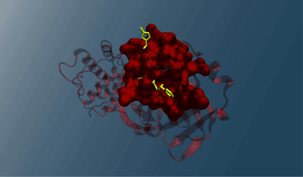
A major therapeutic strategy focuses on bolstering the brain’s natural ability to clear away toxic α-Synuclein aggregates. Immunotherapy is a leading approach, involving the development of monoclonal antibodies and vaccines (αSyn-specific antibodies) that can bind to misfolded α-Synuclein and tag it for removal. Significant research, including work from the Bioinformatics Lab at the Vellore Institute of Technology published in journals like JACS Au, also focuses on rational peptide design to create molecules that can disrupt aggregates, though achieving sufficient CSF penetration remains a challenge. Other strategies, such as Lysosomal Therapeutics, aim to boost the efficiency of the autophagy-lysosomal pathway, effectively re-equipping neurons to dispose of the toxic protein build-up.
Conclusion
The story of α-Synuclein in Parkinson’s disease shows how a protein meant for neuron communication becomes harmful through misfolding, clumping, and cell damage. This change in the original blueprint starts a harmful chain reaction. It causes mitochondrial dysfunction, failed waste removal, and ongoing brain inflammation. This leads to the gradual loss of important dopaminergic neurons. Understanding this process—from the first misfold to widespread brain damage—is key. It not only explains the complex molecular causes of Parkinson’s but also points to new ways to stop it. We can target α-Synuclein directly. We can stop it from clumping, block its spread, or help clear it. This brings us closer to redesigning the blueprint and creating treatments that can stop this disease from getting worse, offering hope for millions affected by this devastating

Leave a Reply