




LATE and Mixed Pathology: Separating Copathology Signals in Blood
Enroll a few hundred older adults with amnestic mild cognitive impairment and a positive amyloid blood test, and a share of them will also carry TDP-43 pathology in the amygdala and hippocampus. Nobody will know which ones. Their decline will be counted as Alzheimer’s decline in both arms, their biomarker trajectories will be read as Alzheimer’s trajectories, and the effect size the trial was powered to detect will shrink by an amount no one can estimate. That is the measurement problem LATE creates.
Can LATE be detected with a blood test?
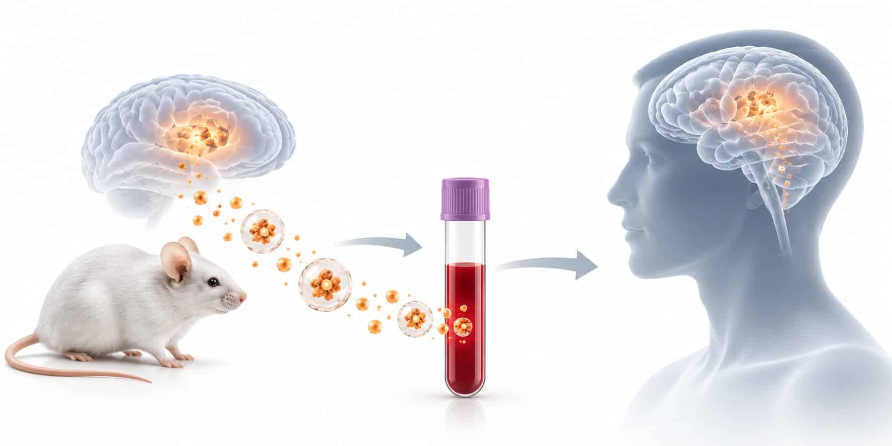
Not with whole-plasma assays alone. Plasma p-tau217 reports Alzheimer’s pathology, while NfL and GFAP report injury and glial response regardless of cause. TDP-43 is expressed throughout the body, so plasma levels mostly reflect peripheral sources. Detecting LATE in blood requires measuring TDP-43 in neuron-derived extracellular vesicles.
Key Takeaways
- LATE neuropathologic change appears in almost 40% of older adults at autopsy and frequently coexists with Alzheimer’s pathology, where the combination produces the steepest cognitive decline.
- Plasma p-tau217 is specific for Alzheimer’s disease by design, so it cannot flag TDP-43 copathology in an amyloid-positive patient.
- NfL and GFAP rise across many neurodegenerative conditions and cannot be partitioned between pathologies in a mixed case.
- TDP-43 in blood is a provenance problem: measuring it inside neuron-derived extracellular vesicles is the most direct route to an attributable, pathology-specific signal.
LATE is the copathology Alzheimer’s trials rarely screen for
Limbic-predominant age-related TDP-43 encephalopathy (LATE) is defined by a stereotyped TDP-43 proteinopathy in older adults, with or without hippocampal sclerosis, and is associated with an amnestic dementia syndrome that mimics Alzheimer’s-type dementia [1]. It is distinguished from frontotemporal lobar degeneration with TDP-43 inclusions by its later age of onset and its relatively restricted neuroanatomical distribution, starting in the amygdala and progressing to the hippocampus and then the middle frontal gyrus [1].
It is not rare. Pooled data from 13 community- and population-based autopsy cohorts, covering 6,196 participants with an average age at death of 88.1 years, found LATE neuropathologic change (LATE-NC) in almost 40% of participants [2]. Among the 3,803 participants with staging data, 25% had LATE-NC above stage 1, the level associated with cognitive impairment, and LATE-NC often, though not always, coexisted with Alzheimer’s disease neuropathologic change (ADNC) [2].
Concurrent pathology produces the worst outcomes
Copathology is not additive noise. In a National Alzheimer’s Coordinating Center analysis of 31 isolated LATE-NC, 244 isolated ADNC and 172 concurrent LATE-NC/ADNC cases, both pathologies impaired cognition in proportion to their stage, and the concurrent group showed the worst overall cognitive effect [3]. Progressive LATE-NC stage carried an independent risk of cognitive impairment (OR 1.66) alongside ADNC level (OR 3.41) [3].
At the individual level, the picture is more fragmented still. In 1,079 autopsied participants from two longitudinal aging studies, 78% had two or more neuropathologies and 35% had four or more; Alzheimer’s pathology was present in 65% but occurred in isolation in only 9% [4]. More than 230 distinct pathology combinations appeared, none in more than 6% of the cohort, and the share of cognitive loss attributable to Alzheimer’s pathology ranged from 22% to 100% across individuals [4].
The pattern holds beyond community cohorts. An analysis of 766 autopsied cases spanning the clinical neurodegenerative spectrum found that older age and APOE ε4 status predicted copathology independently of the primary disease, with direct implications for trials built around monotherapies [5]. LATE-NC also clusters with arteriolosclerosis, hippocampal sclerosis of aging and age-related tau astrogliopathy, and shares genetic risk loci including TMEM106B, GRN, APOE, SORL1 and ABCC9 [6].
What undetected LATE does to a trial
Three distortions follow when TDP-43 copathology goes unmeasured. First, enrollment confuses amyloid-positive with amyloid-driven: a participant can meet every Alzheimer’s biomarker criterion while a meaningful share of their memory loss comes from limbic TDP-43. Second, decline slopes widen. If LATE accelerates decline in some participants in both arms, variance rises and statistical power falls, even with perfect randomization. Third, mechanism-of-action readouts blur. A therapy that clears amyloid cannot be expected to slow TDP-43-driven neurodegeneration, yet downstream injury markers will register both processes as one.
None of this is new to neuropathologists. The 2019 consensus report named the lack of biofluid and neuroimaging biomarkers for antemortem detection as a central obstacle to clinical progress in LATE [1]. What has changed is that trial sponsors now screen on blood, which makes the question of what a blood marker can and cannot see a design decision rather than an academic one.
Why whole-plasma markers cannot attribute the signal
The current generation of plasma biomarkers has earned its place. Plasma p-tau217 has changed amyloid-eligibility screening, and NfL and GFAP are widely used as injury and glial-response readouts. The problem in mixed pathology is not that these markers perform poorly. It is that each one is either specific to one pathology or indifferent to all of them, and neither property helps separate copathology signals.
Plasma p-tau217 is specific, which is why it misses LATE
In 269 older adults from the Religious Orders Study and Rush Memory and Aging Project, with plasma collected an average of 1.4 years before death, plasma p-tau217 correlated more strongly with brain β-amyloid and paired helical filament tau tangles than p-tau181 did [7]. The study tested p-tau against Lewy bodies, TDP-43, hippocampal sclerosis and cerebrovascular pathology, and the authors concluded that the data support p-tau217 specificity for Alzheimer’s disease [7]. A separate autopsy study of 105 participants found plasma p-tau217 and p-tau181 independently associated with plaque and tangle load [8].
That specificity is exactly what an eligibility test should have. It is also why plasma p-tau specificity cannot solve copathology. A marker tuned to Alzheimer’s pathology answers “is Alzheimer’s disease present?” It does not answer “is Alzheimer’s disease the only thing present?” An amyloid-positive participant with advanced LATE-NC and one without it can return comparable p-tau217 values, and the test offers no way to tell them apart.
NfL and GFAP report damage, not its cause
NfL sits at the opposite end. In a multicentre study of 13 neurodegenerative disorders across two cohorts (805 and 1,464 participants), plasma NfL was significantly increased in every cortical neurodegenerative disorder, in ALS and in the atypical parkinsonian disorders [9]. That breadth makes NfL a strong exclusion tool and a weak attribution tool. GFAP behaves similarly: in an autopsy-confirmed cohort spanning Alzheimer’s disease and related dementias, 48% of individuals without ADNC still had increased plasma GFAP [10]. In the head-to-head autopsy comparison, the accuracy of Aβ42/40, GFAP and NfL for predicting Alzheimer’s pathology was lower than that of the p-tau markers [8].
In a patient with concurrent ADNC and LATE-NC, a rising NfL value reflects the sum of both neurodegenerative processes. No arithmetic on that single number recovers the share contributed by TDP-43.
Figure 1. What each blood marker reports in a mixed-pathology cohort
| Marker | What it reports | Pathology-specific? | Can it flag LATE copathology? |
|---|---|---|---|
| Plasma p-tau217 | Amyloid-linked tau pathology | Yes, for Alzheimer’s disease | No — silent on TDP-43 by design |
| Plasma NfL | Axonal injury from any cause | No | No — cannot partition injury by source |
| Plasma GFAP | Astroglial activation | No | No — elevated in many non-Alzheimer’s cases |
| Whole-plasma TDP-43 | Total circulating TDP-43, mostly peripheral | Protein-specific, not tissue-specific | Weakly — brain signal diluted by peripheral pool |
| NDEV TDP-43 | TDP-43 carried in neuron-derived vesicles | Protein- and tissue-specific | Candidate — validation against LATE-NC stage still needed |
Source: synthesized from references [7]–[10] and [12]. The final row reflects evidence from FTD and ALS cohorts; LATE-specific validation is pending.
TDP-43 in blood is a provenance problem, not a sensitivity problem
The obvious fix would be to measure TDP-43 directly in plasma. The obstacle is biological rather than analytical. TDP-43 is a ubiquitously expressed nuclear protein, present in nearly every nucleated cell in the body, including blood cells. A whole-plasma TDP-43 measurement therefore samples a large peripheral pool in which any brain-derived contribution is a small, unlabeled fraction. A more sensitive assay measures that mixed pool more precisely. It does not change where the protein came from.
The pathology itself compounds the problem. LATE is defined by what happens inside neurons: nuclear clearance of TDP-43 and cytoplasmic aggregation. Free circulating protein is an indirect proxy for an intracellular event, and blood-based CNS biomarkers that cannot establish tissue of origin cannot tell a neuronal change from a peripheral one.
The field is pursuing two ways around this. One reads the functional consequence of TDP-43 loss rather than the protein itself: a fluid assay for a cryptic-exon-derived neoepitope detected loss of TDP-43 splicing repression in presymptomatic ALS–FTD [11]. The other restricts measurement to a brain-derived compartment before quantification.
Neuron-derived extracellular vesicles restore the tissue of origin
Extracellular vesicles (EVs) are lipid-bilayer particles that cells release carrying a sample of their cytoplasmic proteins. Neurons release them too, and a fraction of neuron-derived extracellular vesicles (NDEVs) reaches the bloodstream. Enriching plasma for vesicles that display neuronal surface proteins yields a fraction whose cargo reflects neuronal cytoplasm rather than the mixed peripheral pool.
The strongest evidence to date comes from FTD and ALS. In a cohort of 704 patients, including 37 genetically and 31 neuropathologically confirmed cases, plasma EVs contained quantifiable TDP-43 and full-length tau, enabling measurement of 3-repeat and 4-repeat tau isoforms [12]. Most of the EV-associated TDP-43 resided in the neuronally enriched vesicle fraction, and EV 3R/4R tau ratios separated FTLD-tau pathology from FTLD-TDP [12]. In other words, the vesicle compartment carried a pathology-discriminating TDP-43 and tau signal that whole plasma does not.
[IMAGE PLACEHOLDER 2 — scientific schematic]
Filename: ndev-tdp43-provenance-schematic.png
Alt text: Schematic comparing peripheral TDP-43 in whole plasma with neuronal TDP-43 carried in neuron-derived extracellular vesicles
Caption: Whole plasma mixes TDP-43 from every tissue; neuron-derived vesicles carry cargo of known cellular origin.
Visual description: Two-panel schematic. Left panel “Whole plasma”: blood vessel containing many small grey TDP-43 molecules from multiple labeled sources (blood cells, liver, muscle) plus a few blue neuronal molecules. Right panel “NDEV fraction”: neuron at top releasing vesicles into the vessel; vesicles captured by a generic enrichment step (no marker names); vesicle cargo shown as blue TDP-43 and tau. Avoid naming any specific surface marker in the graphic.
A four-axis framework for separating copathology signals
No single analyte resolves mixed pathology. What resolves it is a panel in which each measurement answers one question, and the questions do not overlap. For Alzheimer’s-spectrum programs where LATE is the most likely hidden contributor, four axes cover most of the problem:
- Alzheimer’s axis: plasma p-tau217, answering whether amyloid-linked tau pathology is present.
- Neuronal TDP-43 axis: TDP-43 measured in NDEVs, answering whether neuronal TDP-43 proteinopathy is likely present.
- Injury axis: plasma NfL, answering how much neurodegeneration is occurring from all sources combined.
- Glial axis: plasma GFAP, answering how active the astroglial response is.
Read together, the axes turn one ambiguous cohort into distinguishable subgroups. Where Lewy pathology is also a concern, an NDEV α-synuclein measurement adds a fifth axis on the same draw.
Figure 2. Illustrative biomarker profiles across four copathology subgroups
| Subgroup | p-tau217 | NDEV TDP-43 | NfL | GFAP | Interpretation |
|---|---|---|---|---|---|
| Alzheimer’s only | High | Normal | Elevated | Elevated | Target population for amyloid- or tau-directed therapy |
| Alzheimer’s + LATE | High | Abnormal | Higher | Elevated | Faster expected decline; stratify or adjust |
| LATE, amyloid-negative | Normal | Abnormal | Elevated | Variable | Amnestic syndrome without Alzheimer’s biology; candidate for LATE-directed studies |
| Neither | Normal | Normal | Normal or mild | Variable | Look for vascular, Lewy or non-degenerative causes |
Illustrative. Profiles are conceptual and do not represent measured data or validated cut-points.
Where this changes trial design
The framework earns its cost at four points in a program. At screening, a neuronal TDP-43 readout lets sponsors prespecify a TDP-43-high subgroup instead of discovering heterogeneity after unblinding, which is where most patient stratification failure becomes visible. In the analysis plan, TDP-43 status becomes a baseline covariate in decline models, recovering power lost to copathology-driven variance. For mechanism-of-action claims, it allows a sponsor to ask whether NfL change tracks the targeted pathology or the untargeted one. And for the oldest-old populations where LATE-NC is most prevalent [2], it provides the entry criterion that LATE-directed trials currently lack.
What the evidence supports today, and what it does not
The case rests on three established findings. LATE-NC is common in the age range most Alzheimer’s trials enroll, and its co-occurrence with ADNC worsens cognitive outcomes [2], [3]. Current plasma markers are either Alzheimer’s-specific or pathology-agnostic, so none of them attributes signal to TDP-43 [7], [9]. And the neuronal EV compartment carries quantifiable, pathology-discriminating TDP-43 in FTD and ALS [12].
What remains open is specific. NDEV TDP-43 has not yet been validated against LATE-NC stage in an autopsy cohort with banked antemortem plasma. Cut-points established in FTD and ALS will not transfer directly to an oldest-old population with different baseline biology. Longitudinal stability, pre-analytical handling across sites and the effect of concurrent Alzheimer’s pathology on vesicle cargo all need characterization before the neuronal TDP-43 axis can carry a primary stratification decision. Sponsors evaluating any NDEV assay for a mixed-pathology program should ask for enrichment specificity data, intra- and inter-run CVs, and at least one cohort with neuropathological confirmation.
ExoSORT™ was built for this measurement problem. It enriches plasma for neuron-derived extracellular vesicles so that TDP-43, tau species and α-synuclein are quantified from a brain-origin fraction rather than the mixed peripheral pool. In practice, ExoSORT™ measurements pair with the plasma markers already in a protocol, so one blood draw returns both the Alzheimer’s axis and the neuronal TDP-43 axis.
The next step for the field is the validation study the LATE consensus called for in 2019: antemortem blood, NDEV-based TDP-43 measurement and staged neuropathology in the same participants. Community autopsy cohorts with banked plasma make that study feasible now. Once it exists, LATE can move from an autopsy finding to an enrollment variable, and mixed pathology can be designed into trials rather than discovered in their failures.
References
- [1] Nelson PT, Dickson DW, Trojanowski JQ, Jack CR, Boyle PA, Arfanakis K, et al. Limbic-predominant age-related TDP-43 encephalopathy (LATE): consensus working group report. Brain. 2019;142(6):1503–1527. https://doi.org/10.1093/brain/awz099
- [2] Nelson PT, Brayne C, Flanagan ME, Abner EL, Agrawal S, Attems J, et al. Frequency of LATE neuropathologic change across the spectrum of Alzheimer’s disease neuropathology: combined data from 13 community-based or population-based autopsy cohorts. Acta Neuropathol. 2022;144(1):27–44. https://doi.org/10.1007/s00401-022-02444-1
- [3] Hiya S, Maldonado-Díaz C, Walker JM, Richardson TE. Cognitive symptoms progress with limbic-predominant age-related TDP-43 encephalopathy stage and co-occurrence with Alzheimer disease. J Neuropathol Exp Neurol. 2024;83(1):2–10. https://doi.org/10.1093/jnen/nlad098
- [4] Boyle PA, Yu L, Wilson RS, Leurgans SE, Schneider JA, Bennett DA. Person-specific contribution of neuropathologies to cognitive loss in old age. Ann Neurol. 2018;83(1):74–83. https://doi.org/10.1002/ana.25123
- [5] Robinson JL, Lee EB, Xie SX, Rennert L, Suh E, Bredenberg C, et al. Neurodegenerative disease concomitant proteinopathies are prevalent, age-related and APOE4-associated. Brain. 2018;141(7):2181–2193. https://doi.org/10.1093/brain/awy146
- [6] Nelson PT, Fardo DW, Wu X, Aung KZ, Cykowski MD, Katsumata Y. Limbic-predominant age-related TDP-43 encephalopathy (LATE-NC): co-pathologies and genetic risk factors provide clues about pathogenesis. J Neuropathol Exp Neurol. 2024;83(6):396–415. https://doi.org/10.1093/jnen/nlae032
- [7] Yu L, Boyle PA, Janelidze S, Petyuk VA, Wang T, Bennett DA, et al. Plasma p-tau181 and p-tau217 in discriminating PART, AD and other key neuropathologies in older adults. Acta Neuropathol. 2023;146(1):1–11. https://doi.org/10.1007/s00401-023-02570-4
- [8] Salvadó G, Ossenkoppele R, Ashton NJ, Beach TG, Serrano GE, Reiman EM, et al. Specific associations between plasma biomarkers and postmortem amyloid plaque and tau tangle loads. EMBO Mol Med. 2023;15(5):e17123. https://doi.org/10.15252/emmm.202217123
- [9] Ashton NJ, Janelidze S, Al Khleifat A, Leuzy A, van der Ende EL, Karikari TK, et al. A multicentre validation study of the diagnostic value of plasma neurofilament light. Nat Commun. 2021;12(1):3400. https://doi.org/10.1038/s41467-021-23620-z
- [10] Kivisäkk P, Fatima H, Wu CY, et al. Postmortem associations between Alzheimer disease pathology and plasma pTau217, GFAP, and NfL in AD and AD-related dementias. Neurology. 2026;106(4):e214351. https://doi.org/10.1212/WNL.0000000000214351
- [11] Irwin KE, Jasin P, Braunstein KE, Sinha IR, Garret MA, Bowden KD, et al. A fluid biomarker reveals loss of TDP-43 splicing repression in presymptomatic ALS–FTD. Nat Med. 2024;30(2):382–393. https://doi.org/10.1038/s41591-023-02788-5
- [12] Chatterjee M, Özdemir S, Fritz C, Möbius W, Kleineidam L, Mandelkow E, et al. Plasma extracellular vesicle tau and TDP-43 as diagnostic biomarkers in FTD and ALS. Nat Med. 2024;30(6):1771–1783. https://doi.org/10.1038/s41591-024-02937-4


Leave a Reply