



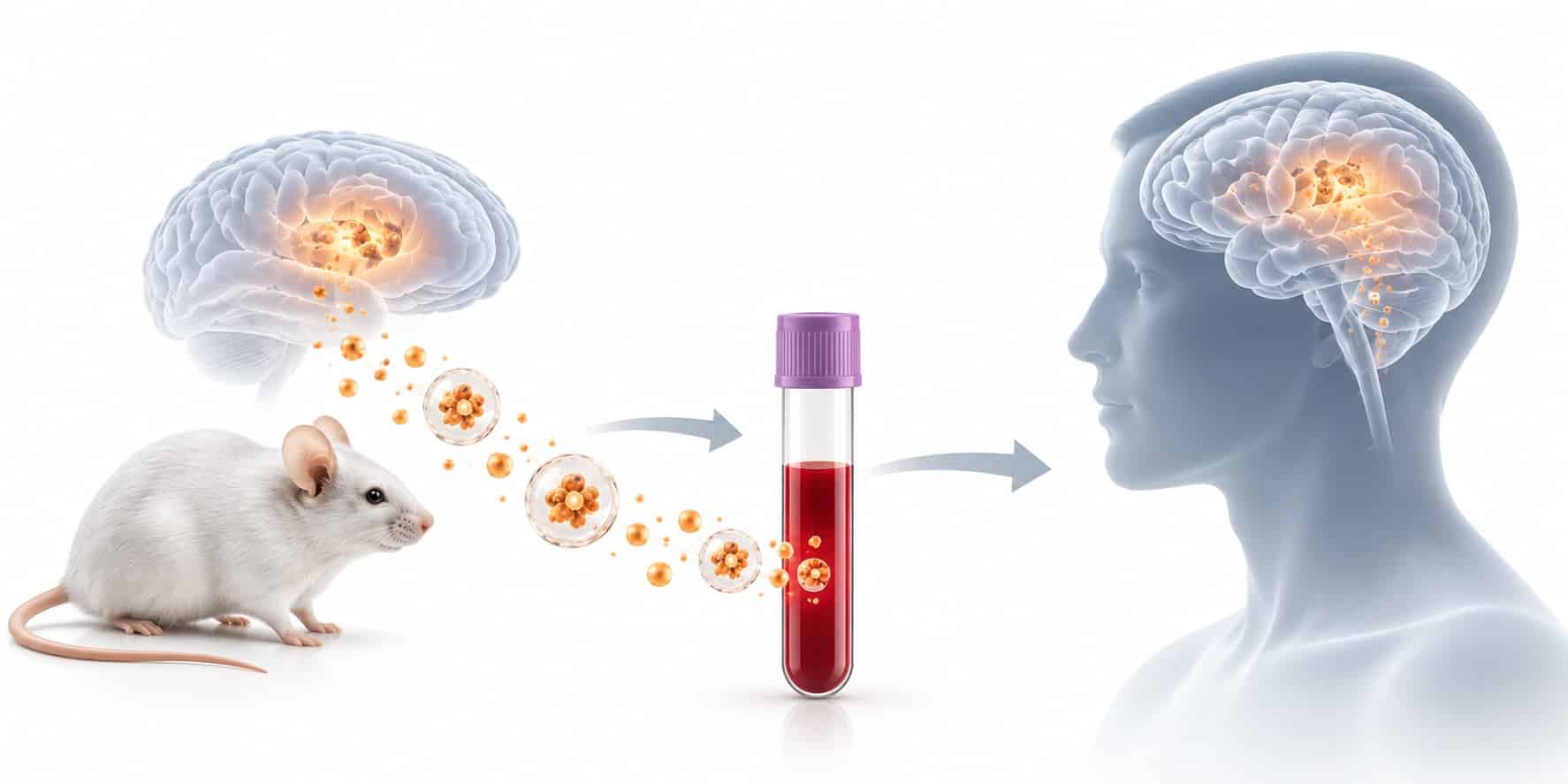
Carrying a Biomarker Signal From Animal Model to First-in-Human
When a research institute re-tested roughly 100 compounds that had looked promising in a widely used ALS mouse model, none of them showed benefit, and eight had already gone on to fail in human trials [1]. The usual lesson drawn from that result is about model design. A second lesson gets less attention. The readouts that told preclinical teams a drug was working rarely had a validated counterpart in human blood. When a preclinical CNS biomarker can’t cross the species boundary, the first-in-human study starts without one.
Key takeaways
- A preclinical CNS biomarker translates only if three things hold steady across species: the analyte, the tissue it comes from, and the assay that measures it.
- Overexpression models amplify brain-derived signal. Human blood dilutes it with peripheral sources, so a whole-plasma marker that looks brain-specific in a mouse may not be in a patient.
- Tissue-of-origin enrichment keeps the measured compartment constant between species, which is what makes a cross-species comparison meaningful.
- A credible bridging package pairs a defined context of use with species-specific analytical validation and blood-to-brain concordance data.
What does it take for a preclinical CNS biomarker to translate to human blood?
A preclinical CNS biomarker translates to human blood when three things carry across species: the same analyte, measured from the same tissue of origin, with an assay validated in each species’ matrix. Blood-to-brain concordance in the model, reproduced in early human sampling, completes the bridge.
The reason this matters goes back to how drug programs fail. An analysis of Phase II decisions across 44 programs at one large sponsor found that most failures came from lack of efficacy, but in 43% of cases the team could not say whether the mechanism had been tested adequately at all [2]. The authors traced survival to three pillars: drug exposure at the site of action, binding to the target, and a measurable pharmacological effect.
For a CNS program, every one of those pillars sits behind the blood-brain barrier. In an animal study you can harvest brain tissue and confirm each one directly. In a human trial you can’t. A blood measurement is the only instrument available at both ends of the program, which makes it the natural bridge, provided it was built to work at both ends from the start. Translational biomarkers that were developed in a mouse and only later adapted to people tend to break at exactly this point.
Why animal-model signals break at the species boundary
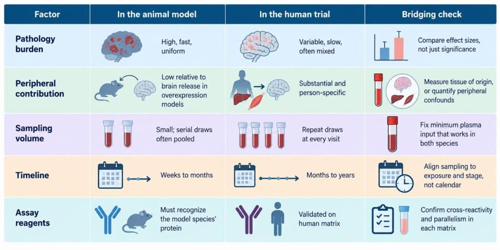
The failure rarely looks dramatic. A marker that separated transgenic animals from wild-type controls with room to spare shows a small, noisy shift in patients, and nobody can tell whether the drug, the disease, or the measurement is responsible. Four mechanisms account for most of that loss.
Pathology burden inflates the dynamic range
Transgenic models are built to produce pathology quickly and at high load. In mouse models of α-synucleinopathy, tauopathy, and β-amyloidosis, plasma neurofilament light chain (NfL) rose between 3- and 100-fold, tracked lesion onset and progression, and correlated closely with CSF levels [3]. NfL is one of the few markers that did make the crossing into human trials, as a measure of neuroaxonal injury. It is also a useful warning. In people, the same analyte sits on top of peripheral modifiers that a mouse cohort never shows: in 662 controls and 2,586 people with multiple sclerosis, plasma NfL fell as body mass index and blood volume rose [4]. Plasma NfL variability of that kind barely registers when the model signal is enormous. It decides the result when the human signal is modest.
Blood volume limits the sampling design
A mouse has very little blood to give. Serial sampling designs that work in a human trial, with a draw at every visit, often can’t be run per animal at all. In one multi-model study of circulating neuronal vesicles, serial samples from each mouse had to be pooled to obtain enough plasma for analysis [5]. That constraint pushes preclinical teams toward terminal endpoints and single timepoints, which in turn leaves the pharmacodynamic time course undefined going into the clinic.
Peripheral background is not the same in both species
A whole-plasma analyte is a sum of every tissue that releases it. Peripheral expression, clearance, and comorbidity differ between an inbred laboratory animal and an aging patient population. A marker whose plasma pool is dominated by brain release in an overexpression model can be dominated by peripheral release in a 70-year-old with reduced kidney function. The analyte name stays the same. What it reports does not.
Disease timelines compress and stretch
Pathology that develops over months in a model develops over years in a patient, and drug exposure profiles differ too. Sampling on matched calendar days rarely corresponds to matched biology. Without a deliberate mapping of exposure and disease stage between species, a marker can look unresponsive simply because it was measured at the wrong moment.
Figure 1. Where a preclinical CNS biomarker loses fidelity between model and patient (illustrative).
Provenance has to translate, too
Every mechanism above has the same root. The measured compartment changes between species, even when the analyte doesn’t. The fix is to hold the compartment constant: measure the analyte from the same cell of origin in the model and in the patient, so that a like-for-like comparison is actually like-for-like.
Extracellular vesicles (EVs), the membrane-bound particles cells release into blood, make that possible because they carry protein and RNA cargo from their parent cells and display parent-cell surface proteins. Pharmacokinetic work in mice has identified a brain-to-blood efflux route that explains how CNS-derived vesicles reach the circulation [6]. Plasma EVs enriched for neuronal origin have been shown to give a more sensitive base for biomarkers than whole plasma, serum, or non-enriched EV preparations [7]. Neuron-derived extracellular vesicles, in other words, give a blood measurement a defined address.
The cross-species evidence is the part that matters most for a bridging study. In a study of 43 mice spanning three Alzheimer’s models and wild-type controls, phosphorylated tau (p181-tau) in circulating neuronal vesicles correlated with levels in cerebral cortex (r = 0.7) and hippocampus (r = 0.6) [5]. That is direct blood-to-brain concordance, the one measurement a human study can’t make outside of autopsy, and it is the anchor a translational team needs before carrying a marker forward. The same requirement applies to biomarker discovery for Alzheimer’s drug programs, where preclinical signals are often the starting point.
The human end of the bridge needs its own evidence. In validation of the ExoSORT™ enrichment method on human plasma, erythrocyte, platelet, and liver mRNA in the enriched fraction fell to under 6% of plasma levels while neuronal mRNA was retained, and repeat processing of the same samples gave coefficients of variation between 8.0% and 22.7% [8]. In a placebo-controlled study, a PPARγ agonist and a GLP-1 receptor agonist each produced drug-specific changes in neuronal-vesicle markers of the Akt–mTOR pathway, and those changes moved largely independently of fasting insulin and glucose [9]. That is a pharmacodynamic readout separating CNS biology from peripheral metabolism in people, which is the job a bridging marker has to do.
One practical caveat. Immunoaffinity capture depends on reagents that recognize the model species’ version of the surface antigen. Before a team designs the animal arm of a bridging study, it should confirm capture and detection reagents cross-react in that species’ plasma, and that the assay shows parallelism in both matrices.
Designing the bridging study: matched matrix, compartment, and timepoints
A bridging study for a preclinical CNS biomarker is not a second discovery study. Its purpose is narrower: to show that a signal observed in the model means the same thing when it is measured in human blood. That purpose dictates the design.
Start with the decision the marker will support in the clinic. A marker intended for CNS target engagement in blood has to move with dose and exposure in the model. A marker intended for pharmacodynamic monitoring in neurodegeneration trials has to show a time course that sampling can resolve. If the marker doesn’t respond to dose in the animal, it won’t serve as a dose-selection readout in humans, no matter how well it separates disease from control.
Then match the pre-analytical chain. Low reproducibility in neurodegeneration fluid biomarker studies has been traced to pre-analytical handling, small samples, and unmeasured confounders [10]. Tube type, time to centrifugation, freeze-thaw history, and storage temperature should be fixed in the animal protocol in a form that a clinical site can reproduce, and EV characterization should be reported to MISEV2023 standards so that both arms are described in the same terms [11].

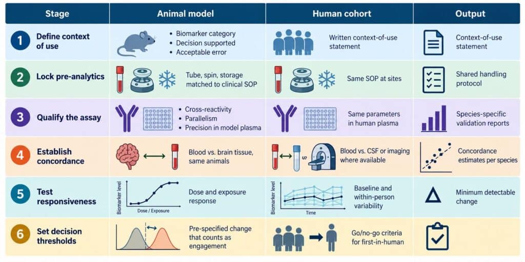
Two design choices are worth making early. The first is plasma input. The minimum volume that the human assay needs should be achievable from a single animal draw, or the study will drift toward pooling and lose per-animal concordance. The second is the human baseline. Within-person variability in the target population sets the smallest change a first-in-human study can detect, and it is cheaper to measure in banked samples before dosing than to discover after a readout comes back ambiguous.
What the bridging data package should contain before IND
Regulators will not ask whether a biomarker is interesting. They will ask what it is for, and whether the method can support that use. The FDA-NIH BEST glossary gives the vocabulary: a marker used to show that a biological response has occurred in someone exposed to a drug is a pharmacodynamic/response biomarker, which is a different claim, with a different evidentiary burden, from a diagnostic or prognostic one [12]. Stating that category up front keeps the package focused.
On the analytical side, FDA guidance on bioanalytical method validation for biomarkers calls for a fit-for-purpose approach and notes that it can also inform methods used on nonclinical study samples [13]. For a bridging marker, that means validating the method in each species’ matrix rather than assuming that performance on human plasma carries over to rodent plasma.
A package that supports a first-in-human decision typically holds six elements: a context-of-use statement; species-specific analytical validation covering precision, parallelism, and sample stability; blood-to-brain concordance from the model; dose or exposure response; baseline and within-person variability in the intended human population; and pre-specified thresholds for what counts as engagement. Programs that carry a preclinical CNS biomarker into the clinic with the first four elements but not the last two are the ones that end up in the 43%: they ran the trial, and they still can’t say whether the mechanism was tested.
The human half of that package is where blood-based CNS biomarkers do most of their work, and it is the half NeuroDex supports. Banked or early-cohort human plasma, processed through ExoSORT™ enrichment, gives a sponsor baseline variability, minimum detectable change, and a pharmacodynamic sampling plan that can be finalized before the first dose.
What comes next: reverse translation
The bridge has mostly been built in one direction, from model to patient. The more useful traffic may run the other way. When a neuronal-vesicle marker moves in patients but not in a given model, that is information about the model, and it should shape which model a program uses for the next compound. Human cohort data, measured from a defined cell of origin, can now inform model selection rather than only validate it after the fact.
That reversal changes what translational biomarkers are for, and what a clinical trial biomarker is for. It becomes a shared reference point that preclinical and clinical teams calibrate against together, rather than a readout handed from one group to the other at IND. For precision neurology, where the same drug may engage its target in one patient and not in the next, a measurement that means the same thing in both species is the starting condition. NeuroDex expects bridging studies built on provenance-matched readouts to become a standard part of IND-enabling work in neurodegeneration within the next few development cycles.
References
- Perrin S. Preclinical research: make mouse studies work. Nature. 2014;507(7493):423-425. https://doi.org/10.1038/507423a
- Morgan P, Van Der Graaf PH, Arrowsmith J, et al. Can the flow of medicines be improved? Fundamental pharmacokinetic and pharmacological principles toward improving Phase II survival. Drug Discov Today. 2012;17(9-10):419-424. https://doi.org/10.1016/j.drudis.2011.12.020
- Bacioglu M, Maia LF, Preische O, et al. Neurofilament light chain in blood and CSF as marker of disease progression in mouse models and in neurodegenerative diseases. Neuron. 2016;91(1):56-66. https://doi.org/10.1016/j.neuron.2016.05.018
- Manouchehrinia A, Piehl F, Hillert J, et al. Confounding effect of blood volume and body mass index on blood neurofilament light chain levels. Ann Clin Transl Neurol. 2020;7(1):139-143. https://doi.org/10.1002/acn3.50972
- Delgado-Peraza F, Nogueras-Ortiz CJ, Volpert O, et al. Neuronal and astrocytic extracellular vesicle biomarkers in blood reflect brain pathology in mouse models of Alzheimer’s disease. Cells. 2021;10(5):993. https://doi.org/10.3390/cells10050993
- Banks WA, Sharma P, Bullock KM, et al. Transport of extracellular vesicles across the blood-brain barrier: brain pharmacokinetics and effects of inflammation. Int J Mol Sci. 2020;21(12):4407. https://doi.org/10.3390/ijms21124407
- Mustapic M, Eitan E, Werner JK Jr, et al. Plasma extracellular vesicles enriched for neuronal origin: a potential window into brain pathologic processes. Front Neurosci. 2017;11:278. https://doi.org/10.3389/fnins.2017.00278
- Eitan E, Thornton-Wells T, Elgart K, et al. Synaptic proteins in neuron-derived extracellular vesicles as biomarkers for Alzheimer’s disease: novel methodology and clinical proof of concept. Extracell Vesicles Circ Nucleic Acids. 2023;4:133-150. https://doi.org/10.20517/evcna.2023.13
- Evers A, Watson K, Abbasi F, et al. Insights from changes in NDEV biomarkers of metabolism: effects of PPARγ and GLP1 receptor agonists on brain metabolism. J Clin Endocrinol Metab. 2026;111(10):2871-2882. https://doi.org/10.1210/clinem/dgag176
- Mattsson-Carlgren N, Palmqvist S, Blennow K, Hansson O. Increasing the reproducibility of fluid biomarker studies in neurodegenerative studies. Nat Commun. 2020;11(1):6252. https://doi.org/10.1038/s41467-020-19957-6
- Welsh JA, Goberdhan DCI, O’Driscoll L, et al. Minimal information for studies of extracellular vesicles (MISEV2023): from basic to advanced approaches. J Extracell Vesicles. 2024;13(2):e12404. https://doi.org/10.1002/jev2.12404
- FDA-NIH Biomarker Working Group. BEST (Biomarkers, EndpointS, and other Tools) Resource. Silver Spring (MD): Food and Drug Administration; Bethesda (MD): National Institutes of Health; 2016-. https://www.ncbi.nlm.nih.gov/books/NBK326791/
- U.S. Food and Drug Administration. Bioanalytical Method Validation for Biomarkers: Guidance for Industry. 2025. https://www.fda.gov/regulatory-information/search-fda-guidance-documents/bioanalytical-method-validation-biomarkers

Leave a Reply