



Plasma p-Tau Specificity in ALS and Alzheimer’s Trials
Plasma p-tau217 has become the reference blood-based CNS biomarker for Alzheimer’s disease, and trial teams now rely on it for screening, enrichment and patient stratification. Plasma p-tau specificity rests on one assumption: that the signal comes from the brain. In ALS, that assumption does not hold. Serum p-tau217 rises in people with no Alzheimer’s pathology, and the evidence points to skeletal muscle [1], [2]. For anyone using blood biomarkers to select patients or read out drug effects, that is a design problem.
Key takeaways
- Serum p-tau217 is elevated in ALS as well as Alzheimer’s disease, while CSF tau is not raised in ALS without Alzheimer’s co-pathology [2].
- Removing peripheral tau before measurement raised discrimination of Alzheimer’s disease from ALS from an AUC of 0.729 to 0.900 [2].
- In muscle, T217 phosphorylation was found only on the peripheral “big tau” isoform [2].
- A blood marker can track clinical progression and still be measuring the wrong tissue.
- Tissue-of-origin evidence belongs in biomarker selection for stratification and pharmacodynamic endpoints.
Why Is Plasma p-Tau217 Elevated in ALS?
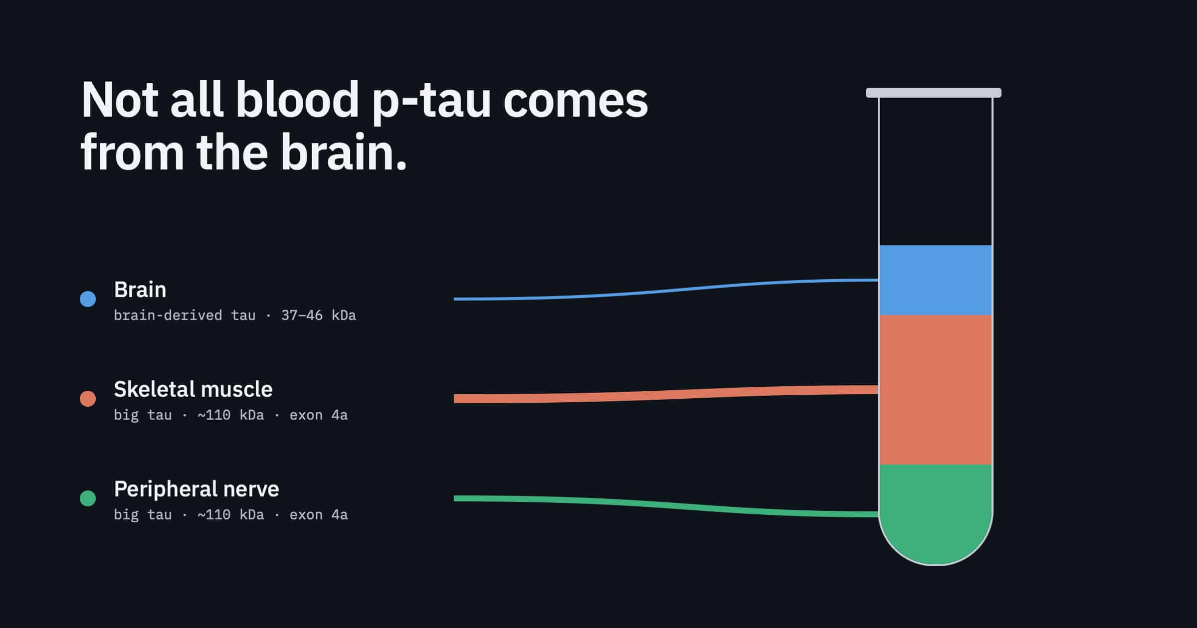
Plasma p-tau217 is elevated in ALS mainly because of tau released from peripheral tissue, not the brain. Skeletal muscle expresses a high-molecular-weight “big tau” isoform that is phosphorylated at threonine 217 and is detected by most commercial p-tau217 immunoassays. CSF tau is not raised in ALS without Alzheimer’s co-pathology, which points away from a CNS source [2].
Blood p-tau assays have earned their place. A 2025 systematic review and meta-analysis concluded that blood phosphorylated tau performs well for diagnosing Alzheimer’s disease across cohorts and assay formats [3]. The weak point is not sensitivity. It is the assumption that every picogram in the tube was shed by a neuron.
The first clear crack came from ALS. In a 2025 multicentre study of 152 people with ALS, 111 with Alzheimer’s disease and 99 disease controls, serum p-tau181 and p-tau217 were higher in both ALS and Alzheimer’s disease than in controls [1]. The same group then looked where nobody had been looking: muscle. Immunohistochemistry and mass spectrometry found both p-tau species in muscle biopsies from ALS patients and controls, with stronger reactivity in atrophic fibres [1].
Assays differ in how strongly they cross-react with that peripheral tau. A head-to-head comparison of p-tau assays found that even the most selective commercial assay still reported moderately raised p-tau217 in ALS compared with controls [4].
Two kinds of tau, two sources
The explanation sits in tau biology. In the CNS, tau is expressed mainly as low-molecular-weight isoforms of roughly 37–46 kDa, often called brain-derived tau. Peripheral tissues mainly express a high-molecular-weight form of about 110 kDa, called big tau, which includes an extra exon (exon 4a) that brain-derived isoforms lack [2]. Big tau expression increases along the path from the central to the peripheral nervous system [5].
That gradient shows up in biofluids. Brain-derived species make up about 99% of total tau in CSF, whereas a substantial fraction of circulating tau in blood is big tau [2]. Most p-tau217 immunoassays target an epitope shared by both forms. A plasma result therefore blends brain and periphery, and in diseases that damage muscle or nerve, the peripheral share grows.
What Isolating Brain-Derived Tau Changed
A study published in Nature Communications in October 2026 tested the peripheral explanation directly [2]. The team measured serum tau in 139 people with ALS, 101 with Alzheimer’s disease and 75 disease controls across four European centres. They compared two approaches:
- One-step: direct measurement with commercial ultrasensitive immunoassays.
- Two-step: a pre-analytical immunoprecipitation using an antibody against the exon 4–exon 5 junction, a sequence present in brain-derived tau but interrupted in big tau, followed by the same immunoassays.
With direct measurement, median serum p-tau217 was 0.37 pg/mL in ALS, 0.75 pg/mL in Alzheimer’s disease and 0.15 pg/mL in controls [2]. ALS sat well above controls despite no Alzheimer’s pathology in most cases.
After brain-derived tau immunoprecipitation in a subset of 149 participants, the picture changed. Brain-derived p-tau217 was elevated only in Alzheimer’s disease, with no significant difference between ALS and controls [2]. Figure 1 shows what that did to diagnostic accuracy.
| Comparison | Direct measurement (AUC) | After brain-derived tau enrichment (AUC) |
|---|---|---|
| Alzheimer’s disease vs. ALS | 0.729 | 0.900 |
| Alzheimer’s disease vs. controls | 0.941 | 0.919 |
| ALS vs. controls | 0.799 | 0.582 |
Two details matter. First, enrichment barely changed the separation of Alzheimer’s disease from controls (0.941 vs. 0.919), which is what you would expect if most p-tau217 in Alzheimer’s disease is already brain-derived. Second, the ALS-vs-control AUC fell to 0.582, close to chance. The earlier “signal” in ALS was largely peripheral. A ratio of brain-derived tau to brain-derived p-tau217 separated Alzheimer’s disease from ALS with an AUC of 0.961 [2].
The tissue evidence
The team also ran targeted mass spectrometry on frontal cortex, spinal cord and muscle. Brain-derived tau dominated in cortex, big tau dominated in muscle, and spinal cord sat in between. When they pulled down p-tau217 from muscle extracts, they detected only big p-tau217 and no brain-derived p-tau217 [2]. In muscle, T217 phosphorylation at measurable levels occurs on the peripheral isoform.
The authors are careful about limits. The tissue analyses used small samples and should be read as supporting mechanistic evidence. Recovery after immunoprecipitation is not yet known, so post-enrichment values are relative rather than absolute. They also note that the two-step method is too complex to serve as a universal first-line test [2].
Peripheral Tau Is Not Just an ALS Problem
ALS makes the confound easy to see because muscle denervation is extensive. The underlying issue applies wherever peripheral tissue is stressed, and trial populations in neurodegeneration are older and often multimorbid. Plasma p-tau specificity therefore depends on who is being tested, not only on the assay.
The Halle group points to several settings where blood p-tau may rise without brain pathology. Elevated blood p-tau217 or p-tau181 has been reported in systemic amyloidosis and myotonic dystrophy type 1. Subclinical muscle and peripheral nerve disease is common and often unrecognised in older adults. Their earlier work also suggested that age- or senescence-related processes may influence p-tau release from muscle independent of disease [2].
Assay labels do not settle the question either. Brain-derived tau assays were designed to target a CNS-enriched epitope, and plasma brain-derived tau has been shown to track amyloid-associated neurodegeneration in Alzheimer’s disease [6], [7]. Yet in the 2026 study, direct measurement with a commercial brain-derived tau assay still read higher in ALS than in controls. The difference only disappeared after selective enrichment [2]. Specificity has to be demonstrated in a population with a known peripheral confound, not inferred from the assay’s design.
What Peripheral Signal Does to Trial Design
For a diagnostic workup, a false positive costs a follow-up test. In a trial, it can cost the study. Peripheral contamination affects the main jobs that blood biomarkers now do in clinical development.
Screening and enrichment
Prevention and early-stage Alzheimer’s trials increasingly use plasma p-tau217 to decide who enrolls. If a share of positive results reflects muscle or nerve rather than brain, the enrolled population is diluted with people who lack the target pathology, and the effect size shrinks. The Halle authors list clinical trials that need high specificity for biomarker-based patient selection, particularly in older or multimorbid populations, among the scenarios where a brain-derived confirmatory step is warranted [2].
Co-pathology in ALS and FTD trials
The reverse problem matters for motor neuron disease programs. Alzheimer’s co-pathology is present in an estimated 10–15% of ALS cases [2]. Standard plasma p-tau217 cannot identify those patients, because most people with ALS already read high. A brain-selective measurement can, which makes it useful for stratification and for interpreting heterogeneous responses.
The correlation trap
This is the most instructive finding for anyone building a pharmacodynamic or progression biomarker. In the ALS cohort, serum brain-derived tau measured directly correlated with ALSFRS-R score (r = −0.471) and with disease progression rate (r = 0.318). After brain-selective enrichment, those associations disappeared [2]. The authors interpret the original correlations as reflecting tau released from degenerating muscle, not neuroaxonal damage.
In other words, a blood marker can track clinical decline convincingly and still be reporting on the wrong tissue. Correlation with disease severity is not evidence of CNS origin. That matters when a sponsor wants to argue that a drug changed brain biology.
Longitudinal monitoring
Change over time is where peripheral contribution is hardest to see. Denervation, muscle mass and renal function can all shift during a trial. The Halle authors note that renal function may influence p-tau217 levels but only minimally affects brain-derived tau [2]. If the peripheral share of a signal drifts while the CNS share stays flat, a longitudinal blood biomarker will report change that has nothing to do with the drug’s mechanism. That is one source of the blood biomarker variability that makes small Phase II trials hard to read.
Tissue of Origin as a Design Requirement
The p-tau story is one well-documented case of a general rule: a blood measurement is only as interpretable as its source. Many proteins that matter in neurodegeneration, TDP-43 among them, are expressed well beyond the brain. Measuring them in bulk plasma raises the same question the ALS data raised for tau.
There are two broad ways to build provenance into a blood-based CNS biomarker:
- Analyte-level selectivity. Target a molecular form that only the CNS produces, as brain-derived tau assays and enrichment steps do [2], [6]. This works when such a form exists and is well characterised.
- Source-level selectivity. Enrich the carriers that neurons release, then measure what they contain. Neuron-derived extracellular vesicles carry cytosolic and membrane proteins from their parent cell, so one enrichment step can serve many analytes, including those with no brain-specific isoform.
Source-level enrichment has its own validation burden, and the field is right to demand it. A 2026 study in the Journal of Extracellular Biology, led by researchers at Columbia University, described an immunoaffinity workflow for enriching nervous-system, lung alveolar and hepatocyte EV subpopulations from human plasma, characterised against MISEV guidance [8], [9]. The neuronal EV fraction showed about a 237-fold enrichment of the synaptic protein synaptophysin over an isotype control and about 482-fold over total plasma EVs [8]. The authors also state plainly that surface markers enrich for tissue-associated EVs rather than prove tissue of origin, and that orthogonal evidence is needed for conclusive assignment [8]. That is the right standard for any provenance claim, brain-derived tau included.
Source-level enrichment has also reached a primary endpoint. In NeuroSense’s Phase 2b trial in ALS (NCT05357950), TDP-43 measured in neuron-derived extracellular vesicles isolated with ExoSORT™ was the primary efficacy endpoint. The company reported a reduction versus placebo at day 180 (p = 0.0421). Patients treated from the start still had lower levels at day 540 than those who began treatment later (p < 0.001) [10]. These are company-reported results from a 68-participant trial, and a larger Phase 3 will determine whether the biomarker change predicts durable benefit. The design point stands regardless: the endpoint was defined by source, not just by analyte.
What to Ask of Any Blood-Based CNS Biomarker
Before a blood marker is used to select patients or support a mechanism claim, these questions are worth answering in writing:
- Is the analyte expressed outside the CNS? If yes, what share of the plasma signal could be peripheral in your population?
- Does the blood signal rise in any condition where CSF does not? A blood-high, CSF-normal pattern, as in ALS for p-tau217, is a strong sign of peripheral origin [2].
- Was specificity tested in a peripheral-confound population? ALS, myopathies and systemic amyloidosis are better tests than healthy controls.
- Do clinical correlations survive tissue-selective measurement? If an association disappears after enrichment, it was not reporting on the brain.
- Could the peripheral share change during the trial? Denervation, muscle mass, age and renal function all move over time.
- For EV-based assays, is enrichment reported to MISEV2023 standards? Size, tetraspanin profile, non-EV contaminants and cell-of-origin validation should all be documented [9].
For most of the last decade, blood biomarker development was a sensitivity race: detect less, earlier. The ALS data show that the next constraint is specificity at the level of tissue. The Halle authors propose a two-level approach, with standard p-tau217 as a first-line test and brain-selective measurement as confirmation where peripheral release could confound results [2]. They also point to quantitative mass spectrometry with labelled reference peptides as a route to absolute measurement of brain-derived tau in biofluids. Expect regulators and trial statisticians to start asking the same question of every blood-based CNS endpoint: not only how low it can detect, but where the signal comes from.
References
- Abu-Rumeileh S, Scholle L, Mensch A, et al. Phosphorylated tau 181 and 217 are elevated in serum and muscle of patients with amyotrophic lateral sclerosis. Nat Commun. 2025;16:2019. https://doi.org/10.1038/s41467-025-57144-7
- Abu-Rumeileh S, Großkopf H, Guthknecht A, et al. Tau isoform mapping across tissues and biofluids reveals a signature distinguishing Alzheimer’s disease from amyotrophic lateral sclerosis. Nat Commun. 2026;17:10745. https://doi.org/10.1038/s41467-026-78390-3
- Therriault J, et al. Blood phosphorylated tau for the diagnosis of Alzheimer’s disease: a systematic review and meta-analysis. Lancet Neurol. 2025;24:740–752. https://doi.org/10.1016/S1474-4422(25)00227-3
- Janelidze S, et al. A comparison of p-tau assays for the specificity to detect tau changes in Alzheimer’s disease. Alzheimers Dement. 2025;21:e70208. https://doi.org/10.1002/alz.70208
- Koppisetti RK, et al. Distribution of big tau isoforms in the human central and peripheral nervous system. Ann Neurol. 2026;100:709–736. https://doi.org/10.1002/ana.78300
- Gonzalez-Ortiz F, et al. Brain-derived tau: a novel blood-based biomarker for Alzheimer’s disease-type neurodegeneration. Brain. 2023;146:1152–1165. https://doi.org/10.1093/brain/awac407
- Gonzalez-Ortiz F, et al. Plasma brain-derived tau is an amyloid-associated neurodegeneration biomarker in Alzheimer’s disease. Nat Commun. 2024;15:2908. https://doi.org/10.1038/s41467-024-47286-5
- Pierri B, Jackson GL, Gololobova O, et al. Immunoaffinity-based protocol to enrich nervous system cell-, lung alveolar cell-, and hepatocyte-derived extracellular vesicles from human plasma. J Extracell Biol. 2026;5(7):e70171. https://doi.org/10.1002/jex2.70171
- Welsh JA, Goberdhan DCI, O’Driscoll L, et al. Minimal information for studies of extracellular vesicles (MISEV2023): from basic to advanced approaches. J Extracell Vesicles. 2024;13(2):e12404. https://doi.org/10.1002/jev2.12404
- NeuroSense Therapeutics. NeuroSense achieves primary endpoint in Phase 2b ALS study with statistically significant reduction of TDP-43, the defining pathological hallmark of ALS [press release]. June 29, 2026. https://www.prnewswire.com/il/news-releases/neurosense-achieves-primary-endpoint-in-phase-2b-als-study-with-statistically-significant-reduction-of-tdp-43-the-defining-pathological-hallmark-of-als-302813175.html

Leave a Reply