



Before You Add a Blood Biomarker to Your Trial, Test It on Samples You Already Have
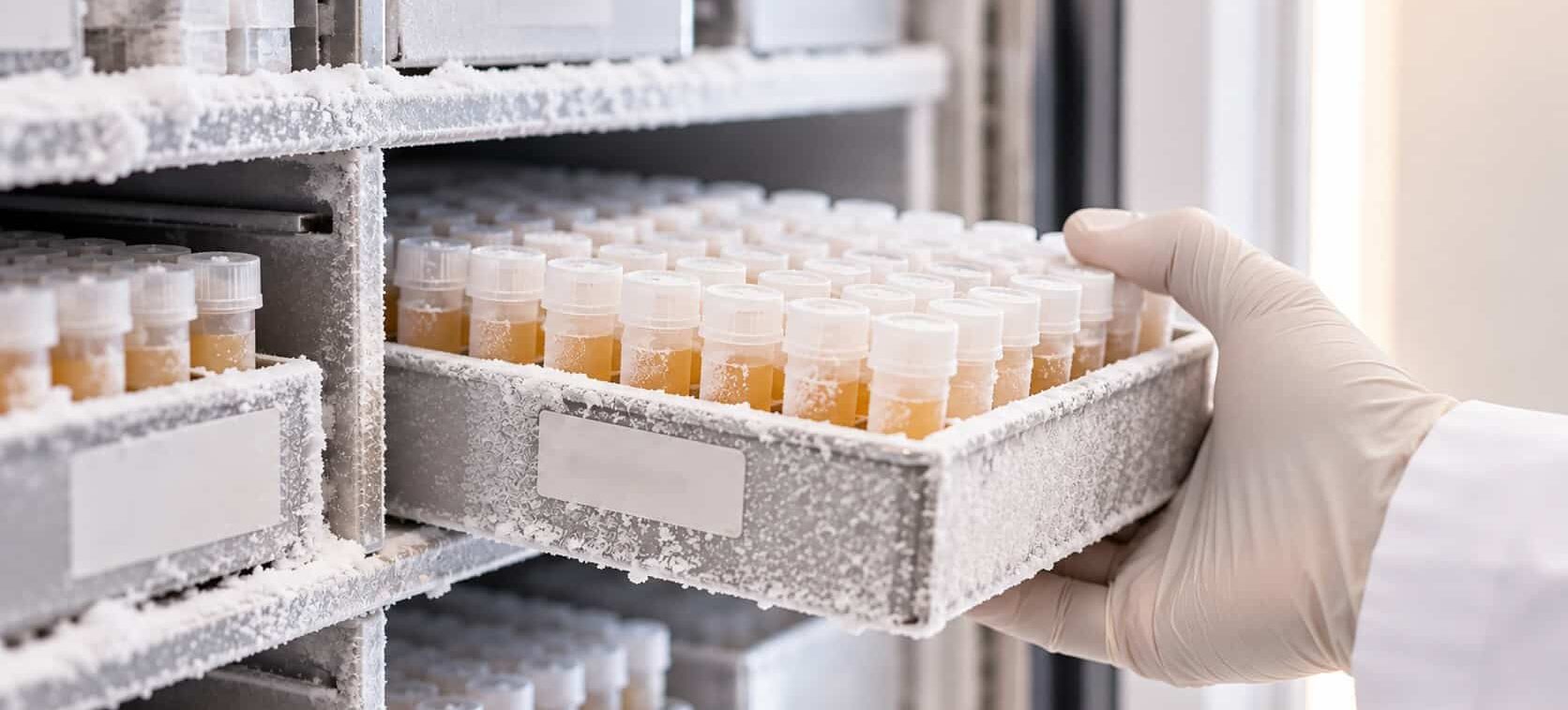
A sponsor writes a blood biomarker into a Phase II protocol on the strength of published cohorts. The marker separated patients from controls in three academic studies, and the assay looked solid. Eighteen months and several hundred enrolled participants later, it fails to separate the sponsor’s own patients. They learned in the trial what they could have learned in a freezer. A feasibility study on banked samples is the cheapest way to find out whether a biomarker will work for your population before your budget depends on it.
Key takeaways
- A banked-sample pilot tests a specific biomarker hypothesis in samples resembling your trial population, before the protocol locks.
- Completed trials, especially their placebo arms, and longitudinal cohorts are often the most valuable sample sources you already have access to.
- Sample history matters: collection tube, processing delays, freeze-thaw cycles, and storage time affect some analytes and EV preparations more than others.
- Set go/no-go criteria, blinding, and sample size before the first sample is thawed, and treat a positive pilot as a reason to validate, not a finished endpoint.
What can a feasibility study on banked samples tell you?
A biomarker feasibility study on banked samples answers four questions before a trial commits to a marker: whether it is measurable in your population, whether it separates the groups your trial depends on, whether it responds to treatment, and how much it varies between draws.
Each question maps to a different failure you would otherwise discover mid-trial. Measurability asks whether the analyte falls inside the assay’s working range in your matrix and population, not only in published cohorts. Separation asks whether the effect size holds in patients who resemble your inclusion criteria, with their age, comorbidities, and disease stage. Treatment response asks whether the marker moves when the mechanism is engaged, which is only testable if treated samples exist. Variability asks how much the marker drifts between repeat draws in the same person, which sets how many participants a trial needs.
This is not discovery research. A feasibility pilot tests a small, pre-specified set of markers against a decision the sponsor has already defined. The distinction matters because the classic phased framework for biomarker development treats retrospective analysis of stored specimens as its own step, before prospective validation. In that framework, the results of earlier phases are generally needed to design the later ones [1]. Skipping the retrospective step does not save time. It moves the learning into the most expensive phase.
Where usable samples come from
Your own completed trials
Completed trials are the most underused biomarker resource most sponsors own. Placebo arms show how a marker behaves over time without intervention: its natural drift, its variability, its relationship to clinical decline. Active arms, if the mechanism is related, show whether the marker responds to drug.
A 2026 study shows how much a re-analysis can deliver. Investigators used ExoSORT™ to enrich neuron-derived extracellular vesicles (NDEVs) from stored plasma from two completed placebo-controlled trials. One tested a PPARγ agonist and the other a GLP-1 receptor agonist. They measured 11 Akt–mTOR pathway proteins at baseline and after 12 weeks. Both drugs produced drug-specific changes compared with placebo, and 77% of participants in the PPARγ agonist group showed an mTOR response [5]. No new participants were enrolled. The samples existed. The question was new.
Longitudinal cohorts
Banked longitudinal samples can answer timing questions that no prospective study could answer quickly. In the TREND study, biennial blood samples from 1,201 people with and without elevated Parkinson’s risk were collected over 4 to 10 years. A later retrospective analysis amplified misfolded α-synuclein from neuronal vesicles in these samples. Every individual who went on to develop Parkinson’s disease tested positive 1 to 10 years before clinical diagnosis, as did 30% of a separate cohort with isolated REM sleep behavior disorder [6]. A prospective study designed to show that would take a decade. The freezer already had it.
Biorepositories and consortia
Disease-focused repositories hold well-annotated plasma from large neurodegeneration cohorts. They are valuable for comparison groups and for indications where a sponsor has no prior trial. Check two things before requesting samples: what clinical and pathological data come attached, and what the access terms allow you to do with results.
Whatever the source, the strongest design follows a principle from biomarker methodology. Specimens should come from a cohort that represents the intended population, collected before outcomes were known, and assayed blind to case status. This prospective-collection, retrospective-blinded-evaluation design removes many of the biases common in biomarker studies [2].
| Sample source | Questions it answers best | Watch out for |
|---|---|---|
| Placebo arm of a completed trial | Natural drift, within-subject variability, link to clinical decline | Consent scope for new analyses; processing SOP of the original trial |
| Active arm of a completed trial | Treatment response, if the mechanism is related | Different mechanism from your drug; small arm sizes |
| Longitudinal natural history cohort | Timing of change, prodromal detection, progression rate | Long storage times; changes in collection protocol across years |
| Disease biorepository | Group separation, comparison and control groups | Mixed sources and handling; limited clinical annotation |
| Pathology- or genetically-confirmed cohort | Whether the marker tracks the underlying pathology | Small numbers; selection toward atypical cases |
What a sample’s history does to the result
A banked sample carries its history with it. The Standardization of Alzheimer’s Blood Biomarkers working group tested common handling variations: collection tube type, delayed centrifugation, centrifugation temperature, aliquot volume, delayed storage, and freeze-thaw cycles. Some analytes were sensitive to these variations while others were resistant. The group used the results to build a standardized plasma handling procedure [7].
The group’s later consensus work found that collection tube and delays before centrifuging or freezing affect Alzheimer’s blood biomarkers. It also found wide differences between analytes: p-tau217 was highly resistant to handling variation, while amyloid-β42 and amyloid-β40 were the most sensitive [8]. Old samples can therefore be fit for one marker and useless for another. Ask for stability data on the specific analyte you plan to test.
Extra considerations for vesicle-based assays
EV preparations add their own storage effects. In a systematic study of eight storage strategies, storage at −80°C reduced EV concentration and sample purity over time, and none of the tested conditions prevented it. Median EV size stayed stable when plasma was stored, but increased when isolated EVs were stored [9]. The practical lesson for banked-sample work is to store plasma, not isolated vesicles, and to record storage duration as a study variable. MISEV2023, the field’s minimal-information guideline, asks researchers to report storage conditions for this reason [10].
Request a sample history before you request samples
For every candidate sample set, ask for:
- collection tube type and anticoagulant;
- time from draw to centrifugation and to freezing;
- storage temperature and any recorded excursions;
- number of prior freeze-thaw cycles;
- collection date and shipping history.
Then check one confound that ruins many retrospective studies. If cases were stored longer than controls, or collected at different sites, storage and site become explanations for any difference you find. Balance groups on storage time and site, or stratify the analysis.
Designing the pilot
Define the decision and the go/no-go criteria first
Write down which trial decision the marker is meant to support, using the FDA-NIH BEST categories: diagnostic, monitoring, prognostic, predictive, pharmacodynamic/response, safety, or susceptibility/risk [4]. Then set thresholds before any sample is thawed. What detection rate, effect size, or response rate would justify carrying the marker forward? Criteria written after the data arrive tend to fit the data.
Size the study for the question
Biomarker sample sizes should follow from three choices: how well a useful marker must perform, how likely the study must be to find it, and how many false leads you can tolerate. In one worked example screening 9,000 candidate markers for cancer recurrence, 40 cases and 160 controls were enough to filter out 98% of useless markers while catching 95% of useful ones [11]. A neurology pilot testing a handful of pre-selected markers needs a different calculation, but it still needs one. A convenience set of 20 samples can only detect very large effects.
Blind the assay and randomize the plates
Run samples blind to group, and interleave cases and controls across plates and runs. If all cases land on one plate, a plate effect becomes a disease effect. Match the validation depth to the pilot’s purpose. An exploratory study does not need the full validation package of a trial endpoint, but precision must be known at the concentrations you expect before any group difference can be interpreted [3].
| Question | What to pre-specify | If it fails |
|---|---|---|
| Measurable? | Minimum proportion of samples within the assay’s quantifiable range | Change matrix, enrichment, or assay before going further |
| Separates groups? | Minimum effect size or AUC in a population like yours | Drop the marker for stratification; it may still serve another role |
| Responds to treatment? | Minimum on-drug vs placebo change, or responder proportion | Do not use as a pharmacodynamic readout |
| Stable enough? | Maximum within-subject variability across repeat draws | Plan more timepoints or more participants, or choose another marker |
Reading the result
A positive pilot justifies a prospective test, not an endpoint. Cut-points derived on small sample sets almost always look better than they will in new data. Lock the cut-point and the analysis plan, then confirm in an independent set or in the trial’s own early samples.
A negative pilot needs a second look before you drop the marker. Separate “the marker does not work” from “these samples could not test it.” Check whether failures cluster by storage time, site, or collection protocol. If they do, the pilot has told you about the samples, not the biology.
Mixed results are often the most useful. A marker that separates groups but varies widely between draws may still work as a stratification tool while failing as a monitoring endpoint.
Whatever the outcome, carry the pilot’s numbers into the protocol. The observed variance sets the power calculation. The timepoints where change appeared set the sampling schedule. The handling conditions that preserved the signal become the trial’s collection SOP. For the timepoint and variability side of that planning, see More Draws, Better Decisions. For the operational side of running samples across many sites, see Turnaround, Batching, and Data Deliverables in a Multi-Site CNS Biomarker Study. And for how a marker moves from pilot to endpoint in ALS specifically, see Three Gates Between an ALS Biomarker and a Trial Endpoint.
Questions to settle with your lab before the pilot starts
What sample volume and type does the pilot need?
Confirm the minimum plasma volume per sample, the acceptable tube types, and whether one aliquot can serve every planned analyte. Volume limits often decide which banked sets are usable at all.
Can the analysis be fully blinded?
The lab should receive coded samples with no group labels, and the unblinding key should stay with the sponsor or a statistician until the pre-specified analysis is run.
Who sets the go/no-go thresholds, and when?
The sponsor should set them, in writing, before samples are thawed. The lab can advise on what is analytically realistic, but thresholds chosen after the data arrive are not thresholds.
What sample metadata does the lab need?
At minimum: collection tube, processing delays, freeze date, freeze-thaw history, storage temperature, and site. Without these, the lab cannot check for handling confounds.
What happens to samples that fail quality control?
Agree in advance on hemolysis limits, minimum volumes, and how failed samples are reported, so that exclusions are documented rather than discovered in the final dataset.
Will the pilot run on the same workflow planned for the trial?
Pilot variance estimates only carry into trial powering if the enrichment, assay, and handling steps match. NeuroDex, for example, scopes engagements to a program’s stage, and a banked-sample pilot can be the first step before trial work.
Trial biobanks are growing faster than anyone is analyzing them. Every completed neurology trial leaves behind placebo-arm plasma with known clinical outcomes, often collected under a defined protocol. As blood-based CNS biomarkers mature, retrospective validation on those samples could become routine before any marker is written into a new protocol. Sponsors who build that step into development planning now will spend less of their trial budget learning what a freezer could have told them.
References
- Pepe MS, Etzioni R, Feng Z, et al. Phases of biomarker development for early detection of cancer. J Natl Cancer Inst. 2001;93(14):1054-1061. https://doi.org/10.1093/jnci/93.14.1054
- Pepe MS, Feng Z, Janes H, Bossuyt PM, Potter JD. Pivotal evaluation of the accuracy of a biomarker used for classification or prediction: standards for study design. J Natl Cancer Inst. 2008;100(20):1432-1438. https://pmc.ncbi.nlm.nih.gov/articles/PMC2567415/
- Lee JW, Devanarayan V, Barrett YC, et al. Fit-for-purpose method development and validation for successful biomarker measurement. Pharm Res. 2006;23(2):312-328. https://doi.org/10.1007/s11095-005-9045-3
- FDA-NIH Biomarker Working Group. BEST (Biomarkers, EndpointS, and other Tools) Resource. Silver Spring (MD): Food and Drug Administration; Bethesda (MD): National Institutes of Health; 2016. https://www.ncbi.nlm.nih.gov/books/NBK326791/
- Evers A, Watson K, Abbasi F, Haque K, Verma A, Eitan E, Rasgon N. Insights from changes in NDEV biomarkers of metabolism: effects of PPARγ and GLP1 receptor agonists on brain metabolism. J Clin Endocrinol Metab. 2026;dgag176. https://doi.org/10.1210/clinem/dgag176
- Kluge A, Schaeffer E, Bunk J, et al. Detecting misfolded α-synuclein in blood years before the diagnosis of Parkinson’s disease. Mov Disord. 2024;39(8):1289-1299. https://doi.org/10.1002/mds.29766
- Verberk IMW, Misdorp EO, Koelewijn J, et al. Characterization of pre-analytical sample handling effects on a panel of Alzheimer’s disease–related blood-based biomarkers: results from the Standardization of Alzheimer’s Blood Biomarkers (SABB) working group. Alzheimers Dement. 2022;18(8):1484-1497. https://doi.org/10.1002/alz.12510
- Verberk IMW, Gouda M, Antwi-Berko D, et al. Evidence-based standardized sample handling protocol for accurate blood-based Alzheimer’s disease biomarker measurement: results and consensus of the Global Biomarker Standardization Consortium. Alzheimers Dement. 2025;e70752. https://doi.org/10.1002/alz.70752
- Gelibter S, Marostica G, Mandelli A, et al. The impact of storage on extracellular vesicles: a systematic study. J Extracell Vesicles. 2022;11(2):e12162. https://doi.org/10.1002/jev2.12162
- Welsh JA, Goberdhan DCI, O’Driscoll L, et al. Minimal information for studies of extracellular vesicles (MISEV2023): from basic to advanced approaches. J Extracell Vesicles. 2024;13(2):e12404. https://doi.org/10.1002/jev2.12404
- Pepe MS, Li CI, Feng Z. Improving the quality of biomarker discovery research: the right samples and enough of them. Cancer Epidemiol Biomarkers Prev. 2015;24(6):944-950. https://doi.org/10.1158/1055-9965.EPI-14-1227


Leave a Reply