



Parkinson’s Starts a Decade Before Diagnosis. Can Blood Find It?
A prevention trial in Parkinson’s disease has a recruitment problem that site activation cannot solve. The people it needs feel well. Most have never seen a movement disorders specialist, and by the time they meet clinical diagnostic criteria, much of the neurodegeneration the drug was meant to prevent is already underway. Two biological frameworks published in 2024 now let researchers define the disease before motor symptoms appear. The open question for drug developers is whether that definition can be applied from an alpha-synuclein blood test.
Key Takeaways
- The NSD-ISS and SynNeurGe frameworks define Parkinson’s disease by biology, not symptoms, which moves diagnostic accuracy upstream into prodromal cohorts [2,3].
- Isolated REM sleep behaviour disorder (iRBD) is the best-characterized prodromal population: 6.3% convert per year, and 73.5% within 12 years [1].
- CSF seed amplification detects misfolded α-synuclein in roughly 85–90% of iRBD patients, but a lumbar puncture does not scale to population screening [5,6].
- Blood assays built on neuron-derived extracellular vesicles have detected seeding-competent α-synuclein up to 10 years before diagnosis, though prodromal sensitivity still trails CSF [9].
- The defensible design today is tiered: blood to screen and track, CSF to confirm a subset.
Can a blood test improve Parkinson’s diagnostic accuracy?
Yes, with qualifications. Blood tests that measure α-synuclein in neuron-derived extracellular vesicles have detected Parkinson’s pathology up to 10 years before clinical diagnosis. In prodromal cohorts they do not yet match CSF seed amplification assays, so the strongest current use is as a scalable screening tier ahead of CSF confirmation and longitudinal tracking.
That answer depends on what Parkinson’s diagnostic accuracy now means. For most of the field’s history, accuracy meant agreement between a clinical diagnosis and autopsy. Since 2024, it increasingly means something harder: detecting the disease process in a person who has no diagnosis at all. The rest of this piece looks at why the target moved, what CSF has established as the benchmark, and where Parkinson’s blood biomarker data currently stand against it.
Can an alpha-synuclein blood test detect Parkinson’s early?
Yes, with qualifications. An alpha-synuclein blood test that measures misfolded α-synuclein in neuron-derived extracellular vesicles has detected Parkinson’s pathology up to 10 years before clinical diagnosis. In prodromal cohorts it does not yet match CSF seed amplification, so its strongest current use is as a scalable screening tier ahead of CSF confirmation and longitudinal tracking.
That answer depends on what Parkinson’s diagnostic accuracy now means. For most of the field’s history, accuracy meant agreement between a clinical diagnosis and autopsy. Since 2024, it increasingly means something harder: detecting the disease process in a person who has no diagnosis at all. The rest of this piece looks at why the target moved, what CSF has established as the benchmark, and where Parkinson’s blood biomarker data currently stand against it.
Biological definitions moved the diagnosis, and the trial population with it
In early 2024, two groups published competing proposals in The Lancet Neurology to define Parkinson’s disease biologically. The Neuronal α-Synuclein Disease Integrated Staging System (NSD-ISS) defines the disease by the presence of pathological neuronal α-synuclein detected in vivo, regardless of clinical syndrome, and adds dopaminergic dysfunction as a second biological anchor [2]. Its earliest stages are asymptomatic: stage 0 is a pathogenic SNCA variant, stage 1A is α-synuclein pathology alone, and stage 1B adds dopaminergic dysfunction [2]. Clinical signs mark the transition to stage 2 and beyond [2].
The SynNeurGe criteria take a three-component approach instead: pathological α-synuclein (S) in tissue or CSF, neurodegeneration (N) on neuroimaging, and pathogenic gene variants (G), each linked to a clinical component [3]. Both groups state that the frameworks are for research use only [2,3]. Both also make the same practical move. They separate the presence of disease from the presence of symptoms.
For trial design, that separation changes the screening question. A clinical diagnosis asks whether a patient’s motor signs fit Parkinson’s disease better than its look-alikes. A biological diagnosis asks whether a person with no motor signs carries the pathology at all. The first question is answered in a clinic. The second has to be answered across thousands of people, most of whom will screen negative, and it has to be answered with a test those people will agree to take.
Neither framework currently names blood as a source for the S component. NSD-ISS was built on CSF seed amplification, and SynNeurGe specifies tissue or CSF [2,3]. That is the gap blood-based biomarkers would need to close, and it is a gap in evidence rather than in principle.
Probabilistic criteria came first
The biological frameworks did not arrive in a vacuum. The Movement Disorder Society (MDS) research criteria for prodromal Parkinson’s disease, updated in 2019, already estimate an individual’s probability of prodromal disease by combining risk markers and prodromal features in a Bayesian model [4]. The update added markers such as diabetes, global cognitive deficit, physical inactivity, and low plasma urate in men, and it explicitly named candidate biomarkers as the route to better prodromal diagnostic accuracy [4]. A validated blood readout of α-synuclein pathology would slot into that model as a high-weight marker, raising or lowering a probability that clinical features alone keep stubbornly uncertain.
iRBD: the best-characterized prodromal cohort, and its screening math
If prevention trials in Parkinson’s disease have a natural starting population, it is isolated REM sleep behaviour disorder. In a multicentre study of 1,280 polysomnography-confirmed iRBD patients across 24 centres, the overall conversion rate to an overt neurodegenerative syndrome was 6.3% per year, with 73.5% converting after 12 years of follow-up [1]. The same analysis estimated that definitive neuroprotective trials in this population would need 142 to 366 patients per arm [1].
Those numbers make iRBD attractive. They also show its limits. Conversion was predicted by abnormal quantitative motor testing, olfactory deficit, mild cognitive impairment, and an abnormal dopamine transporter scan, among other markers [1], which means a sponsor can enrich for near-term converters. But iRBD converts to more than one disease. Patients phenoconvert to Parkinson’s disease, dementia with Lewy bodies, and multiple system atrophy [1]. A synuclein-targeted therapy aimed at neuronal pathology would want to know, at enrollment, which of those trajectories a participant is on.
That is a question of Parkinson’s diagnostic accuracy in its own right, and it is where a cell-type-specific readout earns its place. In a cross-sectional study of 664 serum samples from three European cohorts, α-synuclein in neuron-derived extracellular vesicles was roughly twofold higher in both prodromal (iRBD) and clinical Parkinson’s disease than in multiple system atrophy, controls, or other neurodegenerative diseases [7]. Elevated levels were present before diagnosis and stayed elevated with disease progression [7]. A whole-plasma α-synuclein measurement cannot make that distinction, because it does not know which cells the protein came from. (We cover the reasons in detail in why plasma α-synuclein assays fail Parkinson’s trials.)
| Study | Sample | Measurement | Prodromal population | Key prodromal result |
|---|---|---|---|---|
| Iranzo et al. 2021 [5] | CSF | Seed amplification (RT-QuIC) | iRBD, single centre | 90.4% sensitivity, 90.0% specificity vs. controls |
| Siderowf et al. 2023 [6] | CSF | Seed amplification | iRBD and hyposmia (PPMI) | 44 of 51 positive (28 of 33 with iRBD) |
| Jiang et al. 2020 [7] | Serum | α-synuclein in neuron-derived EVs | iRBD | ~2-fold higher than MSA, controls, other diseases |
| Kluge et al. 2024 [9] | Blood | Seed amplification of neuron-derived EV α-synuclein | Later PD converters; iRBD | All 12 converters positive 1–10 years pre-diagnosis; 30% of 20 iRBD positive |
Source: data as reported in the cited studies [5,6,7,9]. Cohorts, assays, and reference standards differ; results are not directly comparable. Suggested rendering: designer-styled table, four rows, with the blood rows visually grouped.
CSF seed amplification set the benchmark
The reason biological definitions became possible at all is the α-synuclein seed amplification assay (SAA). An SAA uses a small amount of misfolded, seeding-competent α-synuclein in a sample to template aggregation of recombinant protein, amplifying a signal that would otherwise be undetectable. In CSF, its prodromal performance is strong.
In a longitudinal study of iRBD patients, CSF RT-QuIC (real-time quaking-induced conversion, one SAA format) detected misfolded α-synuclein with a sensitivity of 90.4% and a specificity of 90.0%, and a positive result was associated with higher risk of later diagnosis with Parkinson’s disease or dementia with Lewy bodies [5]. The authors proposed CSF SAA positivity as a way to enrich iRBD cohorts in neuroprotective trials [5]. In the multicentre Parkinson’s Progression Markers Initiative (PPMI) cohort, 44 of 51 prodromal participants with iRBD or hyposmia were SAA-positive, including 28 of 33 with iRBD [6].
So CSF answers the biological question well. The difficulty is everything around the answer. A prevention program has to screen a large at-risk population to find the minority who carry pathology, then keep sampling the enrolled participants over years. A lumbar puncture is a reasonable request of a patient with a diagnosis and a treatment decision in front of them. It is a much harder request of a 62-year-old with a sleep complaint, and harder still when the protocol calls for it repeatedly. The bottleneck in prodromal Parkinson’s diagnostic accuracy is not the assay chemistry. It is the matrix.
What neuron-derived extracellular vesicles show in blood today
Blood has always had the access advantage, but an alpha-synuclein blood test has had a provenance problem. Most circulating α-synuclein does not come from the brain, so a total plasma measurement mostly reports peripheral biology. Neuron-derived extracellular vesicles (NDEVs) offer a way around that. Extracellular vesicles are membrane-bound particles that cells release into body fluids, and the subset small enough to originate from the endosomal pathway is often called exosomes. Vesicles carry surface proteins from their cell of origin, so enriching plasma for vesicles of neuronal origin before measurement conditions the result on cell type.
Three studies from one research group now trace how that approach performs across the disease course. In 2022, the group showed that pathological α-synuclein conformers could be detected in NDEVs isolated from plasma, distinguished Parkinson’s patients from individuals without the disease, and could be amplified in a seeding assay [8]. In 2024, the same group ran the blood-based SAA on archived samples from the TREND study, in which 1,201 people with or without elevated Parkinson’s risk gave blood every two years over 4 to 10 years [9]. All 12 participants later diagnosed with Parkinson’s disease were SAA-positive at diagnosis, and all were positive 1 to 10 years before it; healthy controls were negative [9]. A separate study in a larger clinical cohort reported 98.8% sensitivity for diagnosed Parkinson’s disease and found that seeding activity was lower with longer disease duration [10].
That last finding has a practical edge. If NDEV-derived seeding activity is highest early in the disease, the signal is strongest in exactly the window that prevention and early-intervention trials target.
Where blood still trails CSF
The same 2024 study also reported the result a careful sponsor should weigh most heavily. Among 20 iRBD patients from a separate cohort, 30% had a positive blood SAA [9]. CSF studies in iRBD report positivity closer to 85–90% [5,6]. The cohorts differ, the sample is small, and the iRBD group in the blood study had no conversion follow-up reported, so the true sensitivity gap is not yet known. But a gap exists, and a program that treats a negative blood result in an iRBD patient as ruling out synuclein pathology would be overreading the evidence.
Two further cautions apply across this literature. The converter data come from 12 people, which is enough to establish feasibility and not enough to estimate predictive values [9]. And NDEV results depend on how vesicles are isolated and characterized. MISEV2023, the International Society for Extracellular Vesicles reporting guideline, sets the minimum information needed for a reader to judge whether a measured signal genuinely comes from vesicles of the claimed origin [11]. Any NDEV dataset proposed for a trial should be reported against it.
| Study | Sample | iRBD participants | Positive (%) |
|---|---|---|---|
| Iranzo et al. 2021 [5] | CSF | 52 | 90 |
| Siderowf et al. 2023 [6] | CSF | 33 | 85 |
| Kluge et al. 2024 [9] | Blood (neuron-derived EV SAA) | 20 | 30 |
Source: [5,6,9]. Iranzo: 47 positive patients reported as 90% positivity; Siderowf: 28 of 33; Kluge: 30% of 20. Different cohorts and assays; not a head-to-head comparison. Suggested rendering: horizontal bar chart, x-axis “% SAA-positive” 0–100, CSF bars in one colour, blood bar in a second, footnote stating non-comparability.
Designing a prodromal program around a blood readout
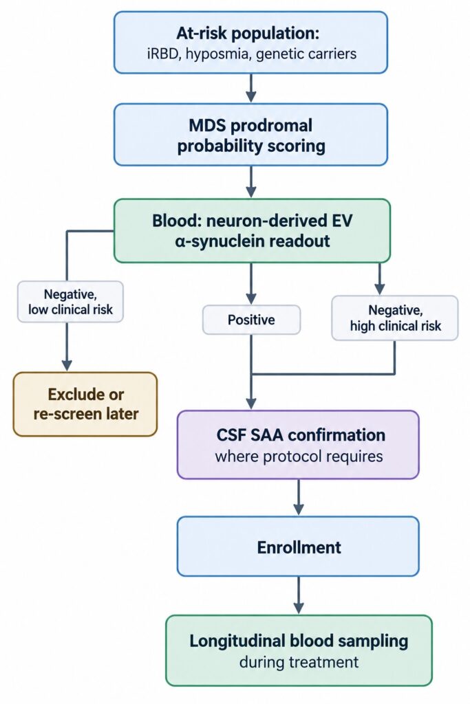
The evidence supports a specific role for blood today, and it is not a CSF replacement. It is the tier that makes the rest of the program affordable. A workable prodromal design has three layers.
Risk enrichment. Start with a population whose prior probability is already elevated: polysomnography-confirmed iRBD, hyposmia, or carriers of pathogenic variants, scored with the MDS prodromal criteria [1,4]. This keeps the number of people who need any biological test manageable.
Blood screening. Apply an alpha-synuclein blood test built on neuron-derived vesicles to the enriched population. Positive results move forward. Given current prodromal sensitivity, negative results in high-risk participants should prompt confirmation rather than exclusion, at least until conversion data clarify what a negative blood result means [9].
CSF confirmation and longitudinal blood sampling. Confirm biological status by CSF SAA in the subset that needs it for eligibility or for regulatory alignment with the NSD-ISS S anchor [2,5]. Then track enrolled participants by blood, as often as the protocol requires, rather than by serial lumbar punctures.
NeuroDex supports this middle tier with ExoSORT™, its platform for isolating neuron-derived extracellular vesicles from plasma, so that α-synuclein and other neuronal cargo can be measured with known cellular provenance. The same question applies to any provider in this space: can they show concordance with CSF SAA in prodromal samples, and can they show it again in an independent cohort?
What to ask for before committing a program
Four questions separate a usable prodromal blood dataset from an encouraging one. First, has the assay been tested in prodromal participants with conversion follow-up, not only in diagnosed patients? Diagnosed-patient sensitivity of 98.8% [10] and iRBD positivity of 30% [9] are both real, and they describe different use cases. Second, is concordance with CSF SAA reported in the same individuals? Third, does the readout distinguish neuronal from oligodendroglial synucleinopathy, given that iRBD can convert to multiple system atrophy [1,7]? Fourth, are isolation and characterization methods reported to MISEV2023 [11]?
Looking ahead: from biological definition to biological screening
For Parkinson’s diagnostic accuracy, the 2024 frameworks did something the field had wanted for decades: they made Parkinson’s disease definable before it is diagnosable [2,3]. What they did not do is make it findable at scale. CSF seed amplification gave the frameworks their foundation, and it will remain the confirmation standard for some time [5,6]. Blood has now shown it can detect the same pathology years before diagnosis [9], but its prodromal sensitivity needs to be measured properly, in larger cohorts with conversion outcomes and paired CSF.
The next few years will likely settle three things: whether an alpha-synuclein blood test built on neuron-derived vesicles can be accepted as a source for the biological S anchor, how their prodromal sensitivity compares with CSF in the same people, and whether they track response to a disease-modifying therapy over time. Sponsors planning prodromal programs now do not have to wait for all three answers. They need a design that uses blood where the evidence is strong and CSF where it is not.
References
- Postuma RB, Iranzo A, Hu M, et al. Risk and predictors of dementia and parkinsonism in idiopathic REM sleep behaviour disorder: a multicentre study. Brain. 2019;142(3):744-759. https://doi.org/10.1093/brain/awz030
- Simuni T, Chahine LM, Poston K, et al. A biological definition of neuronal α-synuclein disease: towards an integrated staging system for research. Lancet Neurol. 2024;23(2):178-190. https://doi.org/10.1016/S1474-4422(23)00405-2
- Höglinger GU, Adler CH, Berg D, et al. A biological classification of Parkinson’s disease: the SynNeurGe research diagnostic criteria. Lancet Neurol. 2024;23(2):191-204. https://doi.org/10.1016/S1474-4422(23)00404-0
- Heinzel S, Berg D, Gasser T, et al. Update of the MDS research criteria for prodromal Parkinson’s disease. Mov Disord. 2019;34(10):1464-1470. https://doi.org/10.1002/mds.27802
- Iranzo A, Fairfoul G, Ayudhaya ACN, et al. Detection of α-synuclein in CSF by RT-QuIC in patients with isolated rapid-eye-movement sleep behaviour disorder: a longitudinal observational study. Lancet Neurol. 2021;20(3):203-212. https://doi.org/10.1016/S1474-4422(20)30449-X
- Siderowf A, Concha-Marambio L, Lafontant DE, et al. Assessment of heterogeneity among participants in the Parkinson’s Progression Markers Initiative cohort using α-synuclein seed amplification: a cross-sectional study. Lancet Neurol. 2023;22(5):407-417. https://doi.org/10.1016/S1474-4422(23)00109-6
- Jiang C, Hopfner F, Katsikoudi A, et al. Serum neuronal exosomes predict and differentiate Parkinson’s disease from atypical parkinsonism. J Neurol Neurosurg Psychiatry. 2020;91(7):720-729. https://doi.org/10.1136/jnnp-2019-322588
- Kluge A, Bunk J, Schaeffer E, et al. Detection of neuron-derived pathological α-synuclein in blood. Brain. 2022;145(9):3058-3071. https://doi.org/10.1093/brain/awac115
- Kluge A, Schaeffer E, Bunk J, et al. Detecting misfolded α-synuclein in blood years before the diagnosis of Parkinson’s disease. Mov Disord. 2024;39(8):1289-1299. https://doi.org/10.1002/mds.29766
- Schaeffer E, Kluge A, Schulte C, et al. Association of misfolded α-synuclein derived from neuronal exosomes in blood with Parkinson’s disease diagnosis and duration. J Parkinsons Dis. 2024;14(4):667-679. https://doi.org/10.3233/JPD-230390
- Welsh JA, Goberdhan DCI, O’Driscoll L, et al. Minimal information for studies of extracellular vesicles (MISEV2023): from basic to advanced approaches. J Extracell Vesicles. 2024;13(2):e12404. https://doi.org/10.1002/jev2.12404



Leave a Reply