



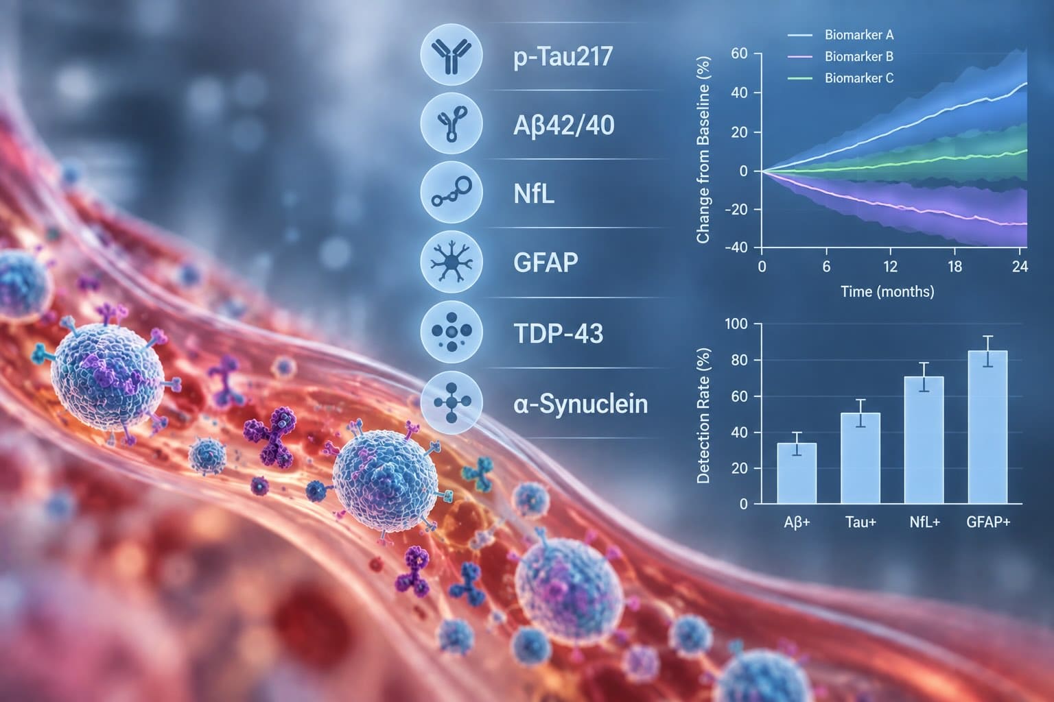
How Blood-Based CNS Biomarkers Improve Trial Endpoints
A sponsor asks the question once a budget line is at stake: if we add blood-based CNS biomarkers to this phase 2, does the trial read out better? It does, and the mechanisms are more specific than the pitch usually suggests. Blood measures do not replace a clinical endpoint. They change the conditions under which that endpoint operates — how much noise it has to overcome, how many participants it needs, and how long the team waits before learning something actionable.
How do blood-based CNS biomarkers improve clinical trial endpoints?
Through four mechanisms. They narrow the variance the endpoint must overcome, remove participants who cannot respond, supply a decision layer that reads out months before the clinical endpoint does, and permit sampling frequent enough to resolve a trajectory rather than a before-and-after comparison. Each requires different evidence, and the last two depend on where the analyte originated.
Key Takeaways
- Biomarker-based screening has cut required sample sizes by 70% in simulated prevention trials, and by 45–60% in enrichment analyses of prodromal cohorts [1].
- Variance reduction improves an existing endpoint rather than replacing it, which is why the gain shows up in power calculations rather than in the endpoint definition.
- Serial CSF collection degrades over time. In a 13-year cohort study, compliance fell from 90% at baseline to 56.2% at year five and 35.7% at year nine [10].
- Pharmacodynamic readouts can carry regulatory weight even without formal qualification — one blood measure supported an accelerated approval in a trial that missed its clinical primary [8,9].
- The last two mechanisms require knowing the analyte came from brain tissue. Mixed-origin plasma concentrations cannot distinguish an ineffective drug from a diluted signal.
Mechanism one: narrowing the variance the endpoint has to overcome
The power of a trial depends on the size of the treatment effect relative to the variability of the outcome. In neurodegeneration, that variability is enormous. Patients carrying the same diagnosis decline at rates differing by several-fold, and that spread sits directly in the denominator of every effect-size calculation. A drug with a real effect can fail to separate from placebo simply because the noise around the endpoint is wider than the signal being sought.
Blood-based markers attack this directly by identifying who is going to decline. In a study of 435 cognitively unimpaired individuals followed for an average of 4.8 years, a screening algorithm combining plasma Aβ42/40, p-tau217 and neurofilament light reduced the sample size required for a simulated trial by 70% when cognition was the endpoint, and by 63% when progression to dementia was the endpoint [1]. Enrichment analyses in prodromal cohorts using markers of amyloid and neurodegeneration together report reductions of 45–60% depending on the endpoint chosen [2].
The same logic holds outside Alzheimer’s, where the enrichment marker differs but the statistical problem does not. ALS trials have historically enrolled broadly and measured function or survival, tolerating wide variation in progression rate. Validated multivariate models now predict individual ALS trajectories from routinely available clinical and demographic variables [7], and blood markers of neuroaxonal damage add prognostic information on top of them. Balancing randomisation across progression strata stops a fast-progressing imbalance in one arm from masquerading as a treatment effect, or from concealing one.
Those numbers describe the same endpoint, measured the same way, in a cohort selected differently. Nothing about the instrument changed. What changed is that the participants enrolled were ones in whom the instrument could register a difference.
This is the cleanest answer to the question of whether a panel improves an endpoint. It improves the endpoint’s operating characteristics, which is what a sponsor actually cares about when they ask whether the trial will read out.
Mechanism two: removing participants who cannot respond
The second mechanism is related but distinct. Variance reduction selects for people who will progress. Cohort purity selects for people who have the pathology the drug targets.
Clinically defined cohorts routinely contain participants whose dominant pathology is not the one under treatment. Seed amplification assays applied to a large, well-characterised Parkinson’s cohort demonstrated substantial biological heterogeneity within a population that met consistent clinical criteria [5]. Every participant enrolled without the target pathology contributes a guaranteed null result. They dilute the treatment effect by construction, and no amount of statistical sophistication recovers what they remove.
Blood-based confirmation changes the economics of that filter. Plasma p-tau217 now performs well enough to triage candidates before confirmatory imaging: in a cross-sectional analysis of 2,916 cognitively unimpaired adults across twelve international cohorts, it achieved 81% accuracy and a positive predictive value of 79% for amyloid status as a stand-alone classifier, positioned by the authors as the first step in a two-step approach that reduces reliance on PET and CSF procedures [3]. The large anti-amyloid programs reporting in 2023 [4,6] each screened substantial populations down to amyloid-confirmed cohorts, and a blood-based first pass changes what that funnel costs without touching the protocol’s endpoint.
Combined with mechanism one, this is where most of the currently demonstrable value sits. It is also where the requirement on the assay is mildest: a screening marker needs to correlate with the pathology of interest, not to prove where it came from.
Mechanism three: a decision layer that reads out before the endpoint does
The first two mechanisms make a clinical endpoint more likely to succeed. The third gives the team information long before that endpoint reports at all.
Clinical endpoints in neurodegeneration are slow by construction. A twelve- to twenty-four-month readout is standard, and in prevention populations the window is longer still. Across that interval a development team is making dose decisions, planning the next study, and allocating capital with no biological confirmation that the molecule is doing anything in the brain. A pharmacodynamic blood measure fills that interval.
The most consequential demonstration came from an antisense oligonucleotide program in a genetic form of ALS. The randomised phase 3 did not meet its primary clinical endpoint, yet treatment produced marked reductions in plasma neurofilament light alongside target-protein reduction in cerebrospinal fluid [8]. The regulator subsequently granted accelerated approval on the basis that the neurofilament reduction was reasonably likely to predict clinical benefit [9]. A blood measure carried a decision the clinical endpoint could not.
Work in multiple sclerosis has explored blood neurofilament light directly as a phase 2 endpoint [12], and biomarker trajectory mapping in familial frontotemporal dementia is beginning to establish when such measures move relative to clinical onset [11] — which is the prerequisite for knowing when to sample.
The honest caveat is that this evidence base is thin. Consensus review of the ALS field noted that support for neurofilament as a response biomarker rested substantially on a single published trial showing meaningful treatment-related change [9]. The mechanism is real and it is demonstrated. It is not yet routine.
Mechanism four: sampling densely enough to see a trajectory
The fourth mechanism is the least discussed and the easiest to quantify.
A biomarker measured twice gives a difference. Measured eight times, it gives a slope, a variance estimate around that slope, and the ability to detect when a trajectory changes. Longitudinal models gain power from repeated measures in a way that no amount of assay refinement substitutes for. The constraint on CNS trials has never been the statistics. It has been access to the compartment.
Serial CSF collection does not hold up across a long study. In a 13-year analysis of 3,479 participants in a major Parkinson’s cohort, compliance with scheduled lumbar punctures fell from 90% at baseline to 76.3% at year one, 67.8% at year three, 56.2% at year five and 35.7% at year nine [10]. Nearly 30% of participants attempting a lumbar puncture experienced an adverse event, most mild, with 1.3% severe [10]. Participant refusal was the most frequently cited reason for unsuccessful collection [10]. This was a research cohort with strong motivation and dedicated infrastructure. An interventional trial in a symptomatic population should not expect to do better.
Missing timepoints are not neutral. They are informatively missing, because the participants who decline a repeat procedure are disproportionately the ones whose disease has progressed. A venous draw is repeatable at every visit, and the resulting series is complete. That difference converts an endpoint measured at two timepoints into one measured across a curve — and a curve detects a divergence between arms considerably earlier than two points do.
What gates the last two mechanisms
The four mechanisms do not make equal demands on the measurement. Mechanisms one and two need correlation. Mechanisms three and four need provenance.
The reason is asymmetric interpretation. For screening, a marker that is partly peripheral in origin still sorts patients usefully, because the question is only whether the pathology is more likely present than absent. For a pharmacodynamic claim, the question is whether a brain-penetrant molecule changed brain biology — and if a large fraction of the circulating analyte never came from brain, a flat result is uninterpretable. The team cannot distinguish a drug that failed from a signal that was diluted. That ambiguity is expensive precisely at the moment the mechanism was supposed to be earning its keep, which is the go/no-go decision.
This is a sample-preparation problem rather than an instrumentation one, and it is the constraint NeuroDex built its platform around. Enriching plasma for neuron-derived extracellular vesicles before measurement establishes tissue of origin at the preparation step instead of inferring it after detection. Vesicles released by neurons carry neuronal cargo into the circulation, so a measurement made inside that compartment reports on brain-derived protein rather than on a pooled plasma concentration of mixed origin. Recent multicohort work using the ExoSORT™ platform measured vesicle-associated α-synuclein across 1,304 individuals in four cohorts with postmortem pathology available for validation [15], which is the scale a response-tier claim requires.
Analyte choice matters alongside compartment. Neurofilament light is a marker of axonal degeneration across conditions rather than of any single disease process [13], and serum concentrations vary substantially with age across the healthy population [14]. A marker that is both non-specific to disease and sensitive to demographics serves mechanisms one and two well and mechanism three only under narrow conditions.
What this builds toward
No blood-based CNS biomarker currently holds formal surrogate endpoint qualification. Since the FDA’s Biomarker Qualification Program opened in 2007, eight biomarkers have completed it, none for a neurological disease [9]. That absence does not prevent a blood measure from influencing a regulatory decision, as the accelerated approval above demonstrates, but it does mean no sponsor should plan a program assuming a purchased panel confers surrogate status. The route there runs through randomised trials that prespecify brain-specific measures and report them whether or not the clinical result cooperates. Programs adding panels now are contributing to that record.
What to ask for before you commit
- Which mechanism are you buying? Variance reduction, cohort purity, earlier decisions, and denser sampling are four different purchases with four different evidence requirements.
- What sample-size reduction does this panel support in your indication? The published figures come from Alzheimer’s cohorts. Ask what the equivalent analysis looks like in yours, and accept an honest answer of “not yet established.”
- How does the measurement establish brain origin? For mechanisms three and four, ask rather than assume. If the answer is that the analyte is mostly brain-derived, ask what fraction and in what population.
- What happens if the marker does not move? Decide before the first sample whether a flat result reads as drug failure or assay limitation. Without a prespecified answer, the panel cannot support a go/no-go decision.
The field’s next few years will be decided in the third and fourth mechanisms rather than the first two, which are largely settled. Enrichment works, the numbers are published, and sponsors are already acting on them. What remains open is whether blood measures can carry mechanism decisions reliably enough to shorten development timelines rather than merely improve the odds on existing ones. That question resolves through compartment-defined measurement and through trials willing to prespecify it — and the answer arrives one randomised dataset at a time.
References
- Cullen NC, Leuzy A, Janelidze S, et al. Plasma biomarkers of Alzheimer’s disease improve prediction of cognitive decline in cognitively unimpaired elderly populations. Nature Communications. 2021;12(1):3555. doi:10.1038/s41467-021-23746-0
- Enrichment of clinical trials in MCI due to Alzheimer disease using markers of amyloid and neurodegeneration. Neurology. 2016;87(12):1235–1241. doi:10.1212/WNL.0000000000003126
- Salvadó G, Janelidze S, Bali D, et al. Plasma phosphorylated tau 217 to identify preclinical Alzheimer disease. JAMA Neurology. Published online September 15, 2025. doi:10.1001/jamaneurol.2025.3217
- van Dyck CH, Swanson CJ, Aisen P, et al. Lecanemab in early Alzheimer’s disease. New England Journal of Medicine. 2023;388(1):9–21. doi:10.1056/NEJMoa2212948
- Siderowf A, Concha-Marambio L, Lafontant DE, et al. Assessment of heterogeneity among participants in the Parkinson’s Progression Markers Initiative cohort using α-synuclein seed amplification: a cross-sectional study. Lancet Neurology. 2023;22(5):407–417. doi:10.1016/S1474-4422(23)00109-6
- Sims JR, Zimmer JA, Evans CD, et al. Donanemab in early symptomatic Alzheimer disease: the TRAILBLAZER-ALZ 2 randomized clinical trial. JAMA. 2023;330(6):512–527. doi:10.1001/jama.2023.13239
- Westeneng HJ, Debray TPA, Visser AE, et al. Prognosis for patients with amyotrophic lateral sclerosis: development and validation of a personalised prediction model. Lancet Neurology. 2018;17(5):423–433. doi:10.1016/S1474-4422(18)30089-9
- Miller TM, Cudkowicz ME, Genge A, et al. Trial of antisense oligonucleotide tofersen for SOD1 ALS. New England Journal of Medicine. 2022;387(12):1099–1110. doi:10.1056/NEJMoa2204705
- Benatar M, Ostrow LW, Lewcock JW, et al. Biomarker qualification for neurofilament light chain in ALS: theory and practice. Annals of Neurology. 2024;95(2):211–216. doi:10.1002/ana.26860
- Barbosa W, Schneider RB, Choi SH, et al. Safety and feasibility of serial lumbar punctures: long-term results from the Parkinson’s Progression Markers Initiative. Clinical Parkinsonism & Related Disorders. 2025;13:100385. doi:10.1016/j.prdoa.2025.100385
- Staffaroni AM, Quintana M, Wendelberger B, et al. Temporal order of clinical and biomarker changes in familial frontotemporal dementia. Nature Medicine. 2022;28(10):2194–2206. doi:10.1038/s41591-022-01942-9
- Sormani MP, Haering DA, Kropshofer H, et al. Blood neurofilament light as a potential endpoint in phase 2 studies in MS. Annals of Clinical and Translational Neurology. 2019;6(6):1081–1089. doi:10.1002/acn3.795
- Khalil M, Teunissen CE, Otto M, et al. Neurofilaments as biomarkers in neurological disorders. Nature Reviews Neurology. 2018;14(10):577–589. doi:10.1038/s41582-018-0058-z
- Simrén J, Andreasson U, Gobom J, et al. Establishment of reference values for plasma neurofilament light based on healthy individuals aged 5–90 years. Brain Communications. 2022;4(4):fcac174. doi:10.1093/braincomms/fcac174
- Gunasekaran TI, Reyes-Dumeyer D, Gu Y, et al. Antemortem α-synuclein levels from neuron-derived extracellular vesicles identify Lewy body pathology in Parkinson’s disease and Alzheimer disease. medRxiv [preprint]. 2026. doi:10.64898/2026.08.03.26359636



Leave a Reply